Category: Blogs

Copy Of Copy Of Copy Of Copy Of Grey Green Modern Corporate Business Presentation 11

How do you diagnose peripheral artery disease?

How Do You Diagnose Peripheral Artery Disease? Exploring Modern Techniques and Expert Insights

Peripheral Artery Disease (PAD) remains a critical health issue, affecting millions of individuals worldwide by impeding blood flow to the limbs. The journey to diagnosing this condition involves a combination of clinical evaluation, symptom assessment, and sophisticated diagnostic tools. In this article, we will delve into how medical professionals diagnose PAD, highlighting the importance of early detection and the latest advancements in medical technology.

Understanding Peripheral Artery Disease

Peripheral artery disease develops when plaque builds up in the arteries that carry blood to the arms and legs, significantly reducing blood flow. Symptoms might include leg pain when walking, leg numbness or weakness, coldness in the lower leg or foot, and sores that do not heal. However, some people with PAD may not exhibit any symptoms, making diagnosis a critical challenge.

Peripheral Artery Disease (PAD) is a common circulatory problem in which narrowed arteries reduce blood flow to your limbs. When you develop PAD, your extremities — usually your legs — don’t receive enough blood flow to keep up with demand. This causes symptoms, most notably leg pain when walking (claudication).

PAD is often a sign of a more widespread accumulation of fatty deposits in your arteries (atherosclerosis). This condition may be reducing blood flow to your heart and brain, as well as your legs.

What is Peripheral Artery Disease?

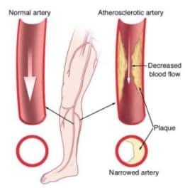
Peripheral artery disease is caused by atherosclerosis, the buildup of plaques made up of fat and cholesterol in the artery walls. As the plaques build up, the arteries become narrowed and stiffened, reducing the flow of oxygen-rich blood to the organs and other parts of the body.

Risk Factors for PAD

Understanding the risk factors for PAD is crucial because managing these can help prevent the condition or mitigate its effects if it develops. Key risk factors include:

– Age: PAD is more common in older adults, typically affecting those over 50.
– Smoking: This is a significant risk factor for PAD. Smoking contributes to the constriction and damage of arteries and raises blood pressure.
– Diabetes: High blood sugar levels associated with diabetes cause damage to blood vessels and also lead to narrowed arteries.
– High cholesterol: Excess cholesterol can contribute to plaque buildup and atherosclerosis.
– High blood pressure: This can damage blood vessels and lead to artery stiffening.
– Obesity: Being overweight is a risk factor for developing PAD.
– Sedentary lifestyle: Lack of exercise contributes to a range of conditions, including heart disease and PAD.
– Family history: A family history of PAD, heart disease, or stroke can increase risk.

Symptoms of Peripheral Artery Disease

The most common symptom of PAD is claudication, which is muscle pain or cramping in the legs or arms that is triggered by activity, such as walking, but disappears after a few minutes of rest. The location of the pain depends on the location of the clogged or narrowed artery. Calf pain is the most common location.

The severity of claudication varies widely, from mild discomfort to debilitating pain. Severe claudication can impede walking and other types of physical activity.

Other symptoms of PAD might include:

– Numbness or weakness in the legs
– Brittle toenails
– Coldness in the lower leg or foot, especially when compared with the other side
– Sores on the toes, feet, or legs that won’t heal
– A change in the color of the legs
– Hair loss or slower hair growth on the feet and legs
– Slower growth of toenails
– Shiny skin on the legs
– Erectile dysfunction, especially in men with diabetes

Complications of Peripheral Artery Disease

If left untreated, PAD can lead to severe health problems, including:

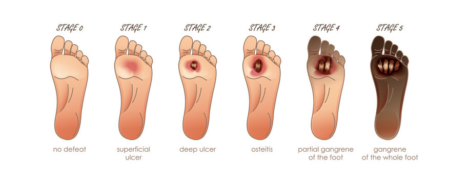
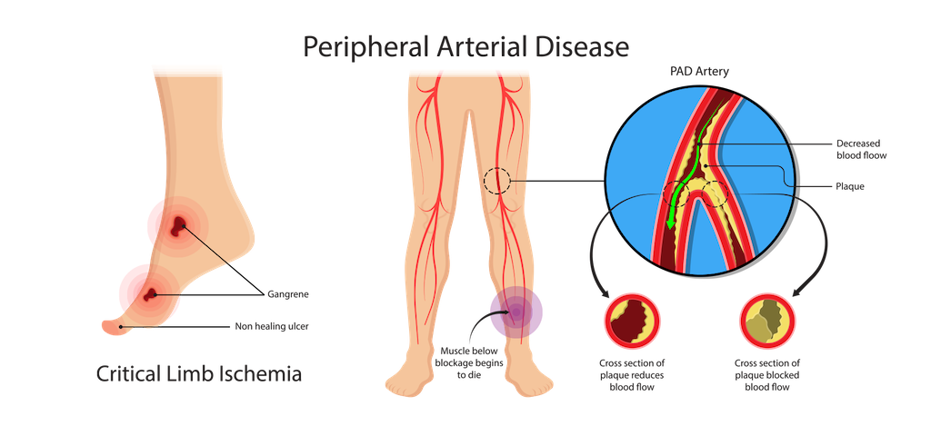
– Critical limb ischemia: This condition begins with open sores that don’t heal, an injury, or an infection of the feet or legs. Critical limb ischemia occurs when such injuries or infections progress and cause tissue death (gangrene), sometimes requiring amputation of the affected limb.
– Stroke and heart attack: The atherosclerosis that causes the signs and symptoms of PAD isn’t limited to your legs. Fat deposits also build up in arteries supplying your heart and brain.

Diagnosis and Treatment

Diagnosis of PAD begins with a physical examination. A key test in diagnosing PAD is the Ankle-Brachial Index (ABI), a simple, non-invasive test in which the blood pressure in the ankles is compared with the blood pressure in the arms. Other imaging tests, such as ultrasound, angiography, and MRI, might be used to highlight the state of the arteries.

Treatment for PAD involves lifestyle changes such as quitting smoking, exercising, and eating a healthy diet. In more severe cases, medication or surgeries such as angioplasty or bypass surgery might be necessary to open up or reroute blood vessels.

Understanding peripheral artery disease is crucial for early detection and treatment, which can significantly improve the quality of life and reduce the risk of complications such as heart attack or stroke. If you suspect you have symptoms of PAD, it is important to consult a healthcare professional for a thorough evaluation and appropriate management.
Peripheral Arterial Disease (Pad) | Cdc.gov

Initial Consultation and Physical Examination

The first step in diagnosing PAD often involves a thorough medical history and physical examination. Doctors will inquire about any symptoms of leg pain, family history of cardiovascular disease, and risk factors such as smoking, diabetes, and hypertension. During the physical exam, the doctor will check for weak pulses in the legs and feet, signs of poor wound healing, and any changes in the color and temperature of the limbs.

Initial Consultation and Physical Examination for Diagnosing Peripheral Artery Disease

When suspecting Peripheral Artery Disease (PAD), the initial consultation and physical examination are crucial first steps in diagnosing the condition. These initial evaluations help healthcare providers not only identify the presence of PAD but also assess the extent and severity of the disease. This stage is vital for developing an effective management plan tailored to the patient’s specific needs.

The Importance of a Detailed Medical History

The first part of the initial consultation involves taking a detailed medical history. This is a critical component as it helps to identify any symptoms that might be indicative of PAD and any risk factors that could predispose the individual to the disease. During this phase, the healthcare provider will ask questions about:

– Symptoms: Inquiring about symptoms such as leg pain or cramps during walking or exercise, which might relieve upon resting, numbness, weakness, or any changes in the color or temperature of the legs.
-Lifestyle habits: This includes smoking history, exercise routines, and dietary habits.
– Medical history: Discussing existing conditions like diabetes, hypertension, high cholesterol, or any family history of cardiovascular disease.
– Medications: Understanding any current medications is important, as some can affect blood flow.

Conducting the Physical Examination

Following the discussion of medical history, the physical examination begins, which is pivotal in observing physical signs that suggest PAD. The examination typically includes several key assessments:

– Pulse measurement: The doctor will check for weak or absent pulses in the legs and feet. Pulse points that are routinely examined include the femoral artery in the groin, the popliteal artery behind the knee, and the dorsal pedis and posterior tibial arteries at the foot.
– Inspection of symptoms and signs: The healthcare provider will look for signs of poor circulation such as shiny skin, color changes, slower hair growth, and ulceration.
– Temperature comparison: Comparing the temperature of different parts of the legs and feet can help identify areas where blood flow might be reduced due to narrowed arteries.
– Capillary refill time: This involves pressing on the toenail or the skin to see how quickly the color returns after being blanched (turned white), which can indicate slowed blood flow.
– Ankle-brachial index (ABI): Although usually part of a more detailed diagnostic procedure, a basic ABI might be performed during the physical examination. This involves measuring blood pressure at the ankle and in the arm to compare the two as a ratio, which can indicate reduced blood flow characteristic of PAD.

What Comes After the Initial Examination?

If the history and physical exam suggest PAD, further diagnostic tests will likely be recommended. These could include more detailed blood flow studies, like duplex ultrasonography, computed tomography angiography (CTA), or magnetic resonance angiography (MRA) to visualize the blood vessels more clearly and identify specific locations and severity of blockages.

In addition to guiding further diagnostic steps, the initial consultation and physical examination help in the risk stratification of the patient. They provide essential data that can be used to tailor interventions, ranging from lifestyle modifications and medications to potentially more invasive procedures like angioplasty or surgery, depending on the severity of the disease.

Peripheral Artery Disease | The Foundation To Advance Vascular Cures

The Ankle-Brachial Index (ABI) Test

A key diagnostic test for PAD is the Ankle-Brachial Index (ABI). This simple, non-invasive test compares the blood pressure in the ankle with the blood pressure in the arm. An ABI ratio of less than 0.90 usually indicates PAD. This test is highly effective as both a screening tool and a diagnostic measure and can be performed in a doctor’s office.

The Ankle-Brachial Index (ABI) Test: A Key Diagnostic Tool for Peripheral Artery Disease

The Ankle-Brachial Index (ABI) test is a simple, non-invasive, and highly effective diagnostic tool used to detect Peripheral Artery Disease (PAD). This test compares the blood pressure in the ankle with the blood pressure in the arm to assess blood flow and identify any blockages or narrowing in the arteries. The ABI test is not only pivotal for diagnosing PAD but also serves as an indicator of the potential risk for other cardiovascular diseases.

How is the ABI Test Performed?

The ABI test is straightforward and can be performed in a healthcare provider’s office with standard blood pressure equipment and a Doppler ultrasound device. The steps involved in conducting the ABI test include:

1. Preparation: The patient is asked to lie flat and rest for about 5 to 10 minutes before the test begins, as resting blood pressure is needed for accurate measurement.
2. Measuring Arm Blood Pressure: Blood pressure cuffs are placed on both arms, and the higher of the two readings is used for the comparison.
3. Measuring Ankle Blood Pressure: The cuffs are then placed on both ankles, and a Doppler device is used to listen to the blood flow in the ankle as the cuff is inflated and then slowly deflated. The pressure at which the pulse is re-detected in the ankle as the cuff deflates is the ankle systolic pressure.
4. Calculation: The ABI is calculated by dividing the higher of the ankle pressures by the higher of the arm pressures.

Interpreting ABI Values

The results of the ABI test are given as a ratio, which can indicate normal blood flow or the presence of PAD:

– Normal ABI: An ABI ratio of 1.0 to 1.4 suggests normal blood flow without significant blockage.
– Mild to Moderate PAD: An ABI ratio of 0.91 to 0.99 may suggest the presence of mild to moderate PAD.
– Severe PAD: An ABI ratio of 0.90 or less is indicative of severe PAD, suggesting significant blockage.
– Above Normal: An ABI ratio greater than 1.4 may indicate rigid arteries and the need for further cardiovascular evaluation, often seen in patients with diabetes.

Benefits of the ABI Test

The ABI test offers several advantages in the clinical setting:

– Non-invasive and Safe: The ABI test involves no injections, no radiation, and is painless, making it a preferred first-line diagnostic tool.
– Cost-effective: Compared to other imaging tests, the ABI test is relatively inexpensive and can be performed quickly.
– Predictive Value: Besides diagnosing PAD, the ABI can indicate a higher risk of cardiovascular events such as heart attacks and strokes, especially in patients with no symptoms.

Limitations and Considerations

While the ABI test is highly valuable, it has some limitations:

– Calcification of Arteries: In patients with severely calcified arteries, such as those with long-standing diabetes, the arteries may not compress when the cuff is inflated, leading to falsely elevated ABI results.
– Operator Dependency: The accuracy of the ABI test can depend on the experience of the healthcare provider performing the test, especially in interpreting Doppler signals.

Doppler and Ultrasound Imaging

For more detailed imaging, doctors may use Doppler and ultrasound technology. These tools help visualize blood flow in the arteries and identify specific locations of blockages. By assessing the flow of blood, these tests not only confirm the diagnosis of PAD but also help in determining the severity of the disease.

Doppler and Ultrasound Imaging: Essential Tools in Diagnosing Peripheral Artery Disease

Doppler and ultrasound imaging represent critical components in the diagnostic process for Peripheral Artery Disease (PAD). These non-invasive imaging techniques provide valuable insights into the blood flow dynamics within the arteries, particularly those in the lower limbs, helping clinicians assess the presence and extent of arterial blockages or narrowing.

Understanding Doppler and Ultrasound Imaging

Ultrasound imaging uses high-frequency sound waves to create images of the inside of the body. When combined with Doppler technology, it not only shows the structure of blood vessels but also measures the movement of blood through these vessels, allowing for the assessment of both anatomical details and functional blood flow information.

How Doppler and Ultrasound Imaging Work

The procedure typically involves the following steps:

1. Preparation: The patient is positioned comfortably, usually lying down, and the area to be examined is exposed.
2. Application of Gel: A clear gel is applied to the skin over the arteries to be examined. This gel helps the ultrasound probe make secure contact with the skin and eliminates air pockets between the probe and the skin surface, which can interfere with sound waves.
3. Probe Placement: The ultrasound technician or doctor moves the probe (transducer) over the area of interest. The probe emits sound waves that bounce off the blood cells moving through the arteries and return to the probe.
4. Image and Sound Wave Analysis: The ultrasound machine processes the sound waves to create images and sounds that can be interpreted by the physician. The Doppler effect allows the device to capture changes in the frequency of the returning sound waves caused by the movement of blood through the arteries.
5. Assessment of Blood Flow: The Doppler signals provide information about the speed and direction of blood flow. Different colors on the Doppler image may indicate the speed and direction of blood flow, which helps identify areas where blood flow is restricted or blocked.

Advantages of Doppler and Ultrasound Imaging

– Non-invasive and Safe: This method does not require incisions or exposure to ionizing radiation, making it safe for repeated use.
– Real-time Results: Doppler and ultrasound imaging provide real-time pictures of blood flow and arterial structure, allowing immediate assessment.
– Detailed Visualization: These techniques can show the thickness of the arterial wall, the presence of plaques, and the severity of narrowing in the arteries.

Applications in PAD Diagnosis

Doppler and ultrasound imaging are particularly useful for:

– Locating Blockages: Identifying the specific locations of arterial blockages or stenosis.
– Assessing Severity: Evaluating the severity of the blockage and how it affects blood flow.
– Guiding Treatment: Assisting in planning medical or surgical treatments, such as angioplasty or bypass surgery.
– Monitoring Progress: Checking the effectiveness of treatments and monitoring progression of the disease over time.

Limitations

While Doppler and ultrasound imaging are highly effective, certain factors can limit their accuracy:

– Operator Skill: The quality of the imaging and the accuracy of the interpretation can depend heavily on the skill and experience of the operator.
– Physical Limitations: In patients with severe obesity or excessive gas in the intestines, obtaining clear images can be challenging.
– Deep Vessel Imaging: Imaging deep vessels or those obscured by bone can sometimes be difficult.
Peripheral Artery Disease | Ctvs Texas

Computed Tomography Angiography (CTA) and Magnetic Resonance Angiography (MRA)

In cases where more detailed images are needed, Computed Tomography Angiography (CTA) and Magnetic Resonance Angiography (MRA) may be utilized. These advanced imaging techniques provide high-resolution images of the arteries, helping pinpoint where arteries are narrowed or blocked.

Treadmill Exercise Test

For patients who can tolerate physical activity, a treadmill exercise test may be recommended. This test helps assess the severity and functional impact of PAD as patients walk until they feel leg pain. Monitoring the walking distance and time helps evaluate the severity of symptoms and effectiveness of potential treatments.

Blood Tests

Blood tests may not directly diagnose PAD, but they are crucial for assessing risk factors and ruling out other conditions. Typical blood tests might include cholesterol levels, triglycerides, diabetes screening, and markers for inflammation such as C-reactive protein (CRP).

Specialist Consultations

In complex cases, or when the diagnosis is uncertain, referral to a vascular specialist may be necessary. These specialists can perform additional tests, such as angiography, where a contrast dye is injected into the arteries before X-rays are taken to view blood flow and pinpoint any blockages.

FAQs on Diagnosing Peripheral Artery Disease

What is the most common symptom of peripheral artery disease?
The most common symptom of PAD is claudication, which manifests as muscle pain or cramping in the legs or arms that occurs during activities such as walking and subsides with rest.

How reliable is the ankle-brachial index test for diagnosing PAD?
The ABI is highly reliable for diagnosing PAD in symptomatic individuals, especially those with typical symptoms like claudication. It is quick, non-invasive, and cost-effective.

Can peripheral artery disease be diagnosed at home?
While PAD cannot be diagnosed at home, individuals can monitor symptoms and risk factors. Professional medical evaluation is necessary for an accurate diagnosis.

Is PAD reversible with medication?
While PAD is not entirely reversible, medications can alleviate symptoms and stop the progression of the disease. Lifestyle changes are also crucial.

How often should someone at risk of PAD be tested?
Individuals at risk of PAD, especially smokers and those with diabetes, high blood pressure, or high cholesterol, should undergo regular screenings as recommended by their healthcare provider.

Conclusion

Diagnosing peripheral artery disease is crucial for managing the disease and improving quality of life. Through a combination of patient history, physical exams, and both basic and advanced diagnostic tests, healthcare providers can effectively identify and manage this condition. Early diagnosis and treatment can significantly reduce the risk of complications, such as heart attack, stroke, and limb amputation. Always consult with a healthcare provider for guidance and the most appropriate testing and management strategies for PAD.

DR. ALOK KUMAR UDIYA

Well-known Interventional Radiologist Dr. ALOK KUMAR UDIYA is currently a consultant at The CARE CHL, an Indore hospital with NABH accreditation. He has a distinguished medical career and has studied at numerous top federal, state, and international superspecialty medical institutes.

He earned his M.B.B.S. from M G M Medical College in Indore and then M.D. in radiodiagnosis from Lady Hardinge Medical College at Delhi University.

Following that, he completed a fellowship in neuro- and vascular interventional radiology at the Sanjay Gandhi Post Graduate Institute of Medical Sciences (SGPGIMS) in Lucknow, where he gained extensive experience in diagnostic imaging along with hepatobiliary, peripheral vascular, and neurovascular interventions.

The prestigious Institute of the liver and biliary sciences Hospital (ILBS), New Delhi, awarded him a P.D.C.C. (Post Doctoral fellowship) in Hepatobiliary intervention, where he also received further in-depth instruction and advanced training in hepatobiliary and transplant imaging and interventions.

Moreover, he completed a six-month Neuro-Interventional Fellowship (FINR) at the famous University of Zurich, where he received specialized training from Professor Anton Valavanis in the endovascular management of stroke and aneurysm. https://cvicvascular.com/

Contact Us

Contact No. –  099993 78980

Email – dralokudiya@gmail.com

Hospital

Care CHL
Address – AB Rd, near L.I.G Square, RSS Nagar, Indore, Madhya Pradesh 452008

Time – 10am to 5pm

Clinic
Address – 403 Panama Tower Geeta Bhawan Square Near Crown Palace, Indore, Madhya Pradesh
Time – 6pm To 8pm

Read More –

Carotid Stenting – https://interventionradiologyindore.com/carotid-stenting/

What Does Early Signs of Gangrene Look Like? – https://interventionradiologyindore.com/what-does-early-signs-of-gangrene-look-like/

What is the cost of AVM embolization in India? – https://interventionradiologyindore.com/cost-of-avm-embolization-in-india/

Copy Of Copy Of Copy Of Copy Of Grey Green Modern Corporate Business Presentation 10

What is the main cause of peripheral vascular disease?

What is the Main Cause of Peripheral Vascular Disease?

Peripheral vascular disease (PVD), also known as peripheral artery disease (PAD), is a common circulatory problem in which narrowed arteries reduce blood flow to the limbs. Understanding the main causes of PVD is crucial for early diagnosis and effective management of the condition. This article delves into the primary cause, explores risk factors, and offers insights into prevention and management strategies for PVD.

Understanding Peripheral Vascular Disease

Peripheral vascular disease primarily affects the blood vessels outside of the heart and brain. The condition is often characterized by a narrowing of the vessels that carry blood to the legs, arms, stomach, or kidneys.

Peripheral Vascular Disease (PVD), also commonly referred to as Peripheral Artery Disease (PAD), involves disorders that cause the blood vessels outside of the heart and brain to narrow, block, or spasm. This can lead to reduced or blocked blood flow, most often in the arteries leading to the lower extremities.

The Physiology and Implications of PVD

The primary physiological change in PVD is the narrowing of peripheral arteries due to atherosclerosis, where plaque builds up on the arterial walls. This plaque is mainly composed of fat, cholesterol, calcium, and other substances found in the blood. Over time, these deposits can harden and reduce the flexibility of the arteries, significantly diminishing blood flow.

Reduced blood flow can lead to several complications:
– Claudication: This symptom involves pain and cramping in the legs due to inadequate blood flow, especially during physical activities.
– Ischemia: Restricted blood flow can cause ischemia, which, if persistent, may lead to tissue damage and necrosis.
– Critical Limb Ischemia (CLI): In severe cases, the lack of blood flow can lead to the development of sores and wounds that do not heal, gangrene, and potentially the need for limb amputation.

Managing and Treating PVD

Management of PVD focuses on reducing symptoms and preventing progression of the disease. This can include:
– Lifestyle Changes: Quitting smoking, exercising regularly, and adopting a heart-healthy diet.
– Medications: Drugs to improve blood flow, manage symptoms, and treat underlying conditions such as high blood pressure and cholesterol.
– Surgical Procedures: In severe cases, procedures such as angioplasty, stenting, or bypass surgery may be required to restore adequate blood flow.

Understanding the complexity of Peripheral Vascular Disease is essential for managing the condition effectively. Early detection and proactive management can help mitigate the severe consequences associated with this disease, improving quality of life and reducing the risk of more severe complications like limb loss.

Peripheral Artery Disease | Ctvs Texas

Atherosclerosis: The Leading Cause of PVD

Atherosclerosis is widely recognized as the principal cause of Peripheral Vascular Disease (PVD), a serious health condition affecting millions globally. This section delves into the mechanisms by which atherosclerosis leads to PVD, its implications, and strategies for management and prevention.

Mechanism of Atherosclerosis

Atherosclerosis begins with damage to the endothelium, the inner lining of the arteries. This damage can be triggered by various risk factors, including high cholesterol, high blood pressure, smoking, diabetes, and inflammatory processes often exacerbated by an unhealthy lifestyle. Once the endothelium is damaged, lipids from the blood can infiltrate the walls of the arteries, leading to the formation of plaques.

These plaques are made up of fats, cholesterol, calcium, and other substances from the bloodstream. Over time, these plaques grow and harden, which narrows the arteries and reduces the elasticity of the vessel walls—a process known as arteriosclerosis. As the artery lumen narrows, the blood flow to various parts of the body becomes restricted, leading to the symptoms and complications associated with PVD.

Impact of Atherosclerosis on Peripheral Arteries

The impact of atherosclerosis on peripheral arteries is significant because these arteries are crucial for supplying blood to the extremities. When plaque buildup narrows these arteries, it directly results in reduced blood flow to the legs and arms. This manifests clinically as claudication—pain and cramping in the affected limbs during activity—due to the muscles not receiving enough oxygen during exertion.

In more severe cases, the reduction in blood flow can lead to critical limb ischemia (CLI), characterized by severe pain at rest, non-healing ulcers, and gangrene. CLI is a serious condition that can result in limb amputation if not adequately addressed.

Understanding the relationship between atherosclerosis and PVD is essential for both patients and healthcare providers to effectively manage this condition. Early diagnosis and proactive management are key to preventing the severe consequences of this disease and maintaining a high quality of life.

Peripheral Artery Disease | The Foundation To Advance Vascular Cures

Risk Factors Contributing to Atherosclerosis and PVD

Risk Factors Contributing to Atherosclerosis and PVD

Atherosclerosis, the underlying cause of Peripheral Vascular Disease (PVD), is a complex process influenced by a variety of risk factors. These risk factors can accelerate the progression of plaque buildup in the arteries, leading to narrowed or blocked arteries which can severely restrict blood flow. Understanding these risk factors is essential for the prevention and management of PVD. Here, we delve into the primary contributors that increase the likelihood of developing atherosclerosis and subsequently PVD.

1. Lifestyle Factors

– Smoking: Smoking is one of the most significant risk factors for atherosclerosis. It damages the lining of the arteries, increases arterial stiffness, raises blood pressure, and reduces the oxygen carrying capacity of the blood, which all contribute to the atherosclerotic process.
– Physical Inactivity: A sedentary lifestyle contributes to the development of atherosclerosis by exacerbating other risk factors such as obesity, high blood pressure, and diabetes.
– Unhealthy Diet: Diets high in saturated fats, trans fats, and cholesterol have been linked to the development of atherosclerosis. These fats contribute to the formation of plaque in the arteries.

2. Medical Conditions

– High Cholesterol Levels: High levels of low-density lipoprotein (LDL) cholesterol (“bad” cholesterol) and low levels of high-density lipoprotein (HDL) cholesterol (“good” cholesterol) can lead to the development of fatty deposits in the blood vessels, which can become atherosclerotic plaques.
– High Blood Pressure: Hypertension can cause damage to the artery walls, making them more susceptible to the accumulation of plaque.
– Diabetes: Diabetes significantly increases the risk of developing atherosclerosis. High blood sugar levels can damage the blood vessel lining and accelerate the growth of atherosclerotic plaque.
– Obesity: Excessive body weight is associated with higher LDL cholesterol levels, higher triglyceride levels, and higher blood pressure, all of which increase the risk of atherosclerosis.

3. Genetic Factors

– Family History: Genetics play a significant role in the development of atherosclerosis. Individuals with a family history of cardiovascular disease, including PVD, are at a higher risk of developing these conditions themselves.

4. Age and Gender

– Age: The risk of atherosclerosis increases with age due to the cumulative effects of exposure to other risk factors and the natural aging of the blood vessels.
– Gender: Men generally have a higher risk of developing atherosclerosis at a younger age compared to women. However, women’s risk increases and may surpass that of men post-menopause.

5. Inflammatory Processes

– Chronic Inflammation: Conditions characterized by chronic inflammation, such as lupus, rheumatoid arthritis, and chronic infections, can also contribute to the development of atherosclerosis. Inflammation plays a direct role in the atherosclerotic process by contributing to the formation and instability of plaques.

Addressing these risk factors not only helps in the prevention of PVD but also mitigates the risk of other cardiovascular diseases, promoting overall vascular health and longevity.

Peripheral Arterial Disease: Causes, Symptoms And Treatment

Symptoms and Diagnosis of PVD

Peripheral Vascular Disease (PVD), also known as Peripheral Artery Disease (PAD), is a common circulatory problem where reduced blood flow due to narrowed arteries leads to various symptoms, primarily in the legs. Recognizing the symptoms and obtaining an accurate diagnosis are crucial steps toward effective management of PVD.

Symptoms of Peripheral Vascular Disease

The symptoms of PVD can vary widely among individuals, and in some cases, the disease may be asymptomatic, particularly in the early stages. The most common symptoms include:

– Claudication: This refers to muscle pain or cramping in the legs or arms that starts during exercise and stops during rest. The pain is typically described as aching, cramping, or inducing fatigue, and it occurs due to inadequate blood flow during increased activity.
– Numbness or Weakness: Reduced blood flow can cause a feeling of numbness or weakness in the legs. This might also be accompanied by a noticeable decrease in temperature in one leg compared to the other.
– Change in Leg Color: As circulation diminishes, there might be noticeable changes in the color of the skin on the legs. The skin may appear paler or take on a bluish tinge.
– Slow Healing Sores: Sores or wounds on the toes, feet, or legs that heal slowly or not at all can be a sign of PVD. This is due to insufficient blood flow needed for healing and tissue repair.
– Hair Loss or Slower Hair Growth: The legs and feet might experience hair loss or reduced hair growth as a result of decreased blood flow.
– Shiny Skin on the Legs: The skin on the legs may look shiny and tight, suggesting changes in the skin due to blood flow issues.
– Pulse Changes in the Legs: The pulses in the feet and legs may become weaker, to the point where they are difficult to detect.

Diagnosis of Peripheral Vascular Disease

Diagnosing PVD involves several steps, typically starting with a review of the patient’s medical history and a physical examination. Key diagnostic tests include:

– Physical Examination: The doctor will look for physical signs of PVD, such as weak or absent pulses in the extremities, reduced blood pressure in the affected limb, or any changes in the appearance of the feet and legs.
– Ankle-Brachial Index (ABI): This simple, non-invasive test compares the blood pressure measured at the ankle with the blood pressure measured at the arm. An ABI ratio of less than 0.9 usually indicates PVD.
– Doppler and Ultrasound Imaging: These imaging tests visualize blood flow in the veins and arteries using high-frequency sound waves. They can show blockages or narrowing of the arteries.
– Angiography: By injecting a contrast agent into the body and taking X-rays, angiography allows for detailed visualization of the blood vessels. This can help identify specific locations and severity of artery narrowing.
– Magnetic Resonance Angiography (MRA) and CT Angiography: These tests provide high-resolution images of the arteries and can show both the blood flow and the structure of the blood vessels.

Prompt diagnosis is essential as PVD can lead to more serious complications, including critical limb ischemia and increased risk of heart attack and stroke. Early diagnosis allows for earlier intervention and better management of the condition to prevent its progression. Lifestyle changes, medications, or in some cases, surgical interventions can significantly improve the quality of life for those affected by PVD.

Prevention and Management of PVD

Preventing and managing Peripheral Vascular Disease (PVD) involves a combination of lifestyle modifications, medical treatments, and in some cases, surgical interventions. The goals are to stop the progression of the disease, alleviate symptoms, improve mobility and quality of life, and reduce the risk of complications such as heart attack, stroke, and limb amputation.

Preventive Measures

Prevention is key in the management of PVD, particularly for individuals at higher risk due to underlying conditions like diabetes, hypertension, or hyperlipidemia. Here are effective strategies to prevent the onset or progression of PVD:

– Quit Smoking: Smoking cessation is crucial, as smoking is a major risk factor for the development and worsening of PVD. Quitting smoking can halt further damage to the arterial walls.
– Regular Physical Activity: Exercise helps improve circulation and heart health, which can prevent the symptoms of PVD from worsening. Walking, cycling, and swimming are excellent forms of aerobic exercise that help condition the muscles to use oxygen more efficiently.
– Healthy Diet: Eating a balanced diet rich in fruits, vegetables, whole grains, and lean proteins can help reduce blood pressure, cholesterol levels, and body weight—all risk factors for PVD.
– Weight Management: Maintaining a healthy weight reduces the burden on the circulatory system and decreases the risk of diabetes and hypertension.
– Manage Blood Sugar Levels: For individuals with diabetes, controlling blood sugar levels is critical to prevent the progression of PVD.
– Regular Health Screenings: Frequent monitoring of blood pressure, cholesterol, and blood sugar levels can help catch and address changes that may indicate an increased risk of PVD.

Management of PVD

Once PVD is diagnosed, management focuses on reducing symptoms and preventing the disease from worsening. The management plan may include:

Medications:
– Antiplatelet agents such as aspirin or clopidogrel to prevent blood clots.
– Cholesterol-lowering drugs (statins) to reduce cholesterol levels and stabilize plaques in the arteries.
– Blood pressure medications to control hypertension.
– Medications for diabetes management to control blood sugar levels.
– Cilostazol or Pentoxifylline to improve symptoms of claudication.

Supervised Exercise Therapy: A structured exercise program supervised by a healthcare professional can significantly improve walking distance and speed for individuals with claudication.

Foot Care: Proper foot care is essential for people with PVD to prevent sores and infections that can lead to serious complications. This includes regular inspection of the feet, wearing appropriate footwear, and keeping the feet clean and moisturized.

Surgical Treatments

For severe cases of PVD where lifestyle changes and medications do not sufficiently alleviate symptoms, surgical options may be considered:

– Angioplasty and Stent Placement: This procedure involves inserting a balloon to expand the narrowed artery, often followed by placing a stent to keep the artery open.
– Bypass Surgery: This involves using a vessel from another part of the body or a synthetic tube to bypass the blocked artery, allowing blood to flow around the narrowed or blocked section.
– Atherectomy: This is a procedure to remove plaque from the arteries.

Ongoing Monitoring and Care

Regular follow-ups with healthcare providers are essential for monitoring the progress of treatment, making necessary adjustments, and ensuring optimal management of the condition. Education about the disease and its management is also crucial for patient compliance and improvement.

Effective prevention and management of PVD are pivotal in mitigating its impact on health and lifestyle. By adopting a proactive approach in addressing risk factors and symptoms, individuals with PVD can lead healthier and more active lives.

Conclusion

The main cause of peripheral vascular disease is atherosclerosis, a condition marked by the buildup of plaque in the arteries. Understanding the risk factors and symptoms associated with this disease is essential for effective management and prevention. By adopting a healthy lifestyle and managing risk factors, individuals can significantly reduce their chances of developing PVD.

FAQs

1. How does smoking affect peripheral vascular disease?
2. What are the best exercises for someone with peripheral vascular disease?
3. What diet changes can help manage or prevent PVD?
4. How often should someone at risk of PVD see a doctor?
5. What are the latest treatments for PVD?
6. How can I tell if I have PVD or another condition like neuropathy?

DR. ALOK KUMAR UDIYA

Well-known Interventional Radiologist Dr. ALOK KUMAR UDIYA is currently a consultant at The CARE CHL, an Indore hospital with NABH accreditation. He has a distinguished medical career and has studied at numerous top federal, state, and international superspecialty medical institutes.

He earned his M.B.B.S. from M G M Medical College in Indore and then M.D. in radiodiagnosis from Lady Hardinge Medical College at Delhi University.

Following that, he completed a fellowship in neuro- and vascular interventional radiology at the Sanjay Gandhi Post Graduate Institute of Medical Sciences (SGPGIMS) in Lucknow, where he gained extensive experience in diagnostic imaging along with hepatobiliary, peripheral vascular, and neurovascular interventions.

The prestigious Institute of the liver and biliary sciences Hospital (ILBS), New Delhi, awarded him a P.D.C.C. (Post Doctoral fellowship) in Hepatobiliary intervention, where he also received further in-depth instruction and advanced training in hepatobiliary and transplant imaging and interventions.

Moreover, he completed a six-month Neuro-Interventional Fellowship (FINR) at the famous University of Zurich, where he received specialized training from Professor Anton Valavanis in the endovascular management of stroke and aneurysm. https://cvicvascular.com/

Contact Us

Contact No. –  099993 78980

Email – dralokudiya@gmail.com

Hospital

Care CHL
Address – AB Rd, near L.I.G Square, RSS Nagar, Indore, Madhya Pradesh 452008

Time – 10am to 5pm

Clinic
Address – 403 Panama Tower Geeta Bhawan Square Near Crown Palace, Indore, Madhya Pradesh
Time – 6pm To 8pm

Read More –

Carotid Stenting – https://interventionradiologyindore.com/carotid-stenting/

What Does Early Signs of Gangrene Look Like? – https://interventionradiologyindore.com/what-does-early-signs-of-gangrene-look-like/

What is the cost of AVM embolization in India? –  https://interventionradiologyindore.com/cost-of-avm-embolization-in-india/

Copy Of Copy Of Copy Of Copy Of Grey Green Modern Corporate Business Presentation 9

What is the cost of AVM embolization in India?

What is the Cost of AVM Embolization in India?

Arteriovenous Malformation (AVM) embolization represents a specialized medical procedure aimed at treating abnormal connections between arteries and veins. This condition, often found in the brain or spine, can lead to serious health complications if untreated. Given the complexity and delicacy of the treatment, AVM embolization requires precise technological expertise and high-level medical skills, which can significantly influence the cost.

In India, the cost of AVM embolization varies widely depending on several factors. This article will explore the price range, contributing elements such as geographical location, hospital facilities, the experience of the medical team, and additional costs that might affect the overall financial planning for patients seeking treatment.

Understanding AVM Embolization

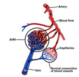
Arteriovenous malformation embolization is a minimally invasive procedure that uses imaging guidance to insert a catheter into the blood vessels to deposit materials that block abnormal blood flow. The primary goal is to reduce the risk of hemorrhage or to alleviate symptoms associated with AVMs.

Avm Embolization Treatment At Best Price In Mumbai | Id: 7982909255

Factors Influencing the Cost of AVM Embolization in India

Factors Influencing the Cost of AVM Embolization in India

Arteriovenous Malformation (AVM) embolization is a crucial medical procedure for patients with abnormal connections between arteries and veins. This detailed look into the factors influencing the cost of AVM embolization in India will help patients and their families understand the financial implications and make informed decisions. The cost varies based on a multitude of factors, from geographical location to the type of technology used.

Geographical Variations**

The location of the hospital significantly impacts the cost of AVM embolization. Hospitals located in metro cities such as Delhi, Mumbai, Chennai, and Bangalore generally have higher costs due to the higher cost of living and operational costs compared to hospitals in smaller cities or rural areas. For example, the price of embolization in a top-tier city hospital can be up to 30-50% higher than in a non-metro hospital.

Hospital Type and Facilities

The type of hospital—private or public—also plays a major role in the cost structure. Private hospitals tend to charge more due to the superior infrastructure, advanced technology, and the availability of specialized care. In contrast, government hospitals usually offer this service at a reduced cost, which can be an advantage for patients looking for more affordable options.

Surgeon’s Expertise and Team Experience

The expertise of the surgeon and the medical team is another critical factor. Experienced neurosurgeons and interventional radiologists who are well-known for their skill in performing complex procedures like AVM embolization often command higher fees. Additionally, a team that includes specialized anesthesiologists, nursing staff, and other support staff who are experienced in pre- and post-operative care can also increase the overall cost.

Type of Technology Used

The cost of AVM embolization is heavily dependent on the type of technology and equipment used. The latest advancements in medical technology, such as digital subtraction angiography (DSA), which provides detailed vascular imaging, can lead to higher costs. Hospitals that invest in state-of-the-art equipment to ensure the highest success rates and lower complications can often have higher procedure costs.

Pre- and Post-Operative Care

Pre-operative care involves various diagnostics and consultations to ensure that the patient is a suitable candidate for the procedure, which can add to the overall cost. Similarly, post-operative care, which may include intensive monitoring and follow-up visits, can also impact the total cost. The extent of care needed varies from patient to patient but is crucial for the procedure’s success and the patient’s recovery.

Insurance and Financial Aid

The extent of insurance coverage is a significant factor for many patients. While most insurance plans in India cover AVM embolization, the level of coverage, terms, and conditions can vary widely between policies and insurers. Patients need to thoroughly understand their insurance benefits and also explore available government aids and subsidies, which can substantially reduce the financial burden.

Additional Costs

There are often additional costs that are not immediately apparent but can affect the overall financial planning for the procedure. These might include transportation, accommodation for post-treatment recovery especially if the patient is from a different city, and any unforeseen complications that may arise necessitating further medical intervention.

Understanding these factors can help patients and their families plan financially for AVM embolization in India. It’s advisable to consult with multiple hospitals and specialists to get a comprehensive view of the expected costs tailored to the specific needs and circumstances of the patient.

Jcm | Free Full-Text | State Of The Art In The Role Of Endovascular  Embolization In The Management Of Brain Arteriovenous Malformations&Amp;Mdash;A  Systematic Review

Average Costs of AVM Embolization in India

Average Costs of AVM Embolization in India

Arteriovenous Malformation (AVM) embolization in India represents a critical treatment option for patients dealing with this complex condition. Understanding the financial aspects of AVM embolization is vital for patients and their families as they prepare for potential medical expenses. The cost of this procedure in India can vary widely based on numerous factors, but here we’ll provide a clearer picture of the average expenses involved.

Range of Costs Across India

The cost of AVM embolization in India can range significantly depending on the complexity of the case, the hospital, and the region. On average, patients might expect to pay anywhere from ₹1,50,000 to ₹4,50,000. This range reflects both the minimum and maximum costs that one might encounter in various settings across the country.

Breakdown of Procedure Costs

1. Initial Consultation and Diagnostic Tests: Before the embolization procedure, patients must undergo thorough diagnostic tests including MRI, CT scans, and angiography to determine the specifics of the AVM. These tests can cost between ₹10,000 to ₹50,000, depending on the hospital and the type of imaging used.

2. Procedure Costs: The actual embolization procedure is the most significant expense. The cost includes the use of specialized equipment and materials, such as catheters and embolic agents, and the fees for the medical team performing the operation. This can range from ₹1,00,000 to ₹3,50,000.

3. Hospital Stay and Post-Operative Care: Post-procedure, a hospital stay of one to several days may be necessary, depending on the patient’s recovery. The cost for this stay, which includes nursing care, bed charges, and meals, can add another ₹20,000 to ₹50,000 to the bill.

Comparison with International Costs

Comparing these costs with those in Western countries illustrates a significant cost advantage in India. For instance, in the United States, the cost of AVM embolization can range from $20,000 to $50,000, making India a cost-effective destination for those seeking high-quality medical care at a fraction of the price.

Insurance and Financing Options

Most health insurance policies in India cover the cost of AVM embolization, but the extent of coverage can vary. It’s essential for patients to consult with their insurance provider to understand the specifics of what is covered and what out-of-pocket expenses might remain. Additionally, many hospitals in India offer financing options that allow for the payment to be spread out over time, making the procedure more accessible.

Additional Financial Considerations

Patients should also account for indirect costs such as travel, accommodation for family members, and any potential loss of income during recovery. These costs, while not directly related to the medical procedure, can add up and should be considered when planning for treatment.

Seeking the Best Value

Patients are advised to seek detailed quotations from several hospitals and to inquire about all possible expenses to avoid unexpected costs. Consulting with health care professionals and planning extensively can help manage the financial impact of AVM embolization effectively.

In conclusion, while the cost of AVM embolization in India is considerably lower than in many Western countries, it is still a significant financial undertaking. Understanding these costs allows patients to make informed decisions and arrange the necessary funds, ensuring they receive the care they need without undue financial strain.

Avm Treatments In India And Hyderabad | Dr. Suresh Giragani

Patient Experiences with AVM Embolization in India

Many patients from within India and abroad have reported positive experiences with AVM embolization in Indian hospitals, noting the high level of care and expertise available.

Conclusion

The cost of AVM embolization in India is influenced by a variety of factors including the location, type of hospital, technology used, and the expertise of the medical team. While affordable compared to Western standards, patients considering this treatment should carefully research and plan their medical journey to ensure they receive the best possible care at a reasonable cost.

FAQs

1. How long does recovery from AVM embolization take?
2. Are there any risks associated with AVM embolization?
3. What are the symptoms that indicate I might need AVM embolization?
4. How do I choose the right hospital for AVM embolization in India?
5. Can international patients access AVM embolization services in India?
6. How effective is AVM embolization in preventing future health issues?

DR. ALOK KUMAR UDIYA

Well-known Interventional Radiologist Dr. ALOK KUMAR UDIYA is currently a consultant at The CARE CHL, an Indore hospital with NABH accreditation. He has a distinguished medical career and has studied at numerous top federal, state, and international superspecialty medical institutes.

He earned his M.B.B.S. from M G M Medical College in Indore and then M.D. in radiodiagnosis from Lady Hardinge Medical College at Delhi University.

Following that, he completed a fellowship in neuro- and vascular interventional radiology at the Sanjay Gandhi Post Graduate Institute of Medical Sciences (SGPGIMS) in Lucknow, where he gained extensive experience in diagnostic imaging along with hepatobiliary, peripheral vascular, and neurovascular interventions.

The prestigious Institute of the liver and biliary sciences Hospital (ILBS), New Delhi, awarded him a P.D.C.C. (Post Doctoral fellowship) in Hepatobiliary intervention, where he also received further in-depth instruction and advanced training in hepatobiliary and transplant imaging and interventions.

Moreover, he completed a six-month Neuro-Interventional Fellowship (FINR) at the famous University of Zurich, where he received specialized training from Professor Anton Valavanis in the endovascular management of stroke and aneurysm. https://cvicvascular.com/

Contact Us

Contact No. –  099993 78980

Email – dralokudiya@gmail.com

Hospital

Care CHL
Address – AB Rd, near L.I.G Square, RSS Nagar, Indore, Madhya Pradesh 452008

Time – 10am to 5pm

Clinic
Address – 403 Panama Tower Geeta Bhawan Square Near Crown Palace, Indore, Madhya Pradesh
Time – 6pm To 8pm

Read More –

Brain Stroke – https://interventionradiologyindore.com/brain-stroke/

Revolutionizing Treatment: The Benefits and Risks of Aneurysm Coiling Unveiled”Aneurysm Coiling- https://interventionradiologyindore.com/aneurysm-coiling/

What Does Early Signs of Gangrene Look Like? – https://interventionradiologyindore.com/what-does-early-signs-of-gangrene-look-like/

Copy Of Copy Of Copy Of Copy Of Grey Green Modern Corporate Business Presentation 8

What Does Early Signs of Gangrene Look Like?

Gangrene, a serious medical condition that involves the death of body tissue due to a lack of blood supply or severe infection, can progress quickly if not identified and treated early. Recognizing the early signs of gangrene can significantly improve the prognosis by facilitating prompt treatment. This article delves into what these early signs look like, the underlying causes, and effective early detection strategies.

Introduction to Gangrene

Understanding Gangrene and Its Implications

Gangrene is not just a single condition but a serious symptom of underlying health issues that can lead to severe consequences, including amputation or even death, if untreated. It is crucial to understand its early symptoms to prevent its progression. Gangrene is a severe medical condition that occurs when a significant mass of body tissue dies due to a lack of blood supply or severe infection. This condition can affect any area of the body but most commonly involves the extremities like toes, fingers, limbs, and organs. Gangrene can develop rapidly and requires immediate medical attention to prevent life-threatening complications, including the need for amputation or severe systemic infection.

Initial Symptoms of Gangrene

The early signs of gangrene typically include a noticeable change in skin color to red, which then progresses to brown and eventually black. The area may feel cool to the touch due to the lack of blood flow and might be accompanied by pain or numbness. As the condition progresses, the symptoms become more pronounced, with the skin potentially breaking down and emitting a foul odor if an infection is present.

Color Changes

– Pale to Blue/Black Transition: One of the first noticeable signs of gangrene is a noticeable change in skin color. Affected areas may turn from red to brown and eventually to black.
– Skin Temperature Changes: The affected area might feel cooler compared to the surrounding skin due to the reduced blood flow.

Sensory Changes

– Numbness and Pain: Early gangrene can cause a range of sensations from pain to numbness, depending on the type and location of the affected tissue.

Early Diagnose Of Gangrene Symptoms Can Aid Its Treatment

Types of Gangrene and Early Signs of Gangrene

There are primarily two types of gangrene: dry and wet

Dry Gangrene

Dry gangrene arises from a reduction of blood flow through the arteries and is more common among people with vascular diseases, such as diabetes and atherosclerosis. The affected tissue typically appears dry, shriveled, and dark brown or black. Unlike wet gangrene, dry gangrene develops slowly and is not usually accompanied by infection, making it less immediately dangerous but still serious.

Characteristics and Early Signs: This type of gangrene typically begins with a dry and shriveled appearance of the skin which changes color to dark brown or black. Dry gangrene usually occurs in the extremities like toes and fingers.

Wet Gangrene

Wet gangrene, on the other hand, occurs when the body tissue becomes infected with bacteria. It can develop after a severe burn, frostbite, or injury that causes a loss of blood supply to the affected area. The presence of infection causes the tissue to swell and emit a foul odor. This type of gangrene spreads much quicker than dry gangrene and can be fatal if not treated promptly.

– Detection and Symptoms: Often resulting from an infection in an injury or surgical wound, wet gangrene may present with swelling, blistering, and a wet appearance. The skin may also emit a foul odor.

Gas Gangrene

A particularly dangerous form of wet gangrene is gas gangrene, which is caused by a specific bacterium known as Clostridium perfringens. This bacterium produces gas within the tissue, which can be felt as a crackling sensation when the area is pressed. Gas gangrene is extremely aggressive and can spread rapidly through the body, making swift medical intervention critical.

– Early Indications: One of the most dangerous types, gas gangrene, can develop rapidly, producing gas within tissue, which can be detected by a crackling sensation under the skin.

Risk Factors and Causes

Contributing Conditions

– Diabetes and Vascular Diseases: These conditions are significant risk factors because they affect blood circulation, particularly in the extremities.
– Injuries and Infections: Any severe injury or infection that decreases blood supply to an area can lead to gangrene.

Diagnosing Gangrene in Early Stages

The diagnosis of gangrene involves a physical examination, during which a doctor will observe the color and temperature of the affected area. Imaging tests, such as X-rays, CT scans, or MRIs, can assess the extent of the condition. Blood tests may also be used to detect infection or other related conditions like diabetes.

Medical Assessment and Imaging

– Physical Examination: Early gangrene diagnosis often starts with the visible symptoms on the skin.
– Imaging Tests: Techniques such as MRI or CT scans may be employed to assess the extent and location of the gangrene.

Gangrene Images – Browse 5,208 Stock Photos, Vectors, And Video | Adobe Stock

Preventive Measures

Preventing gangrene involves managing any underlying conditions that increase the risk of developing it, such as diabetes or peripheral arterial disease. Regular examination of the feet and hands for signs of injury or infection is particularly important for those at higher risk. Maintaining a healthy lifestyle, such as quitting smoking and eating a balanced diet, also helps improve blood circulation.

Lifestyle Adjustments

– Managing Diabetes: Proper management of diabetes is crucial in preventing gangrene. Regular monitoring of blood sugar levels and maintaining a healthy lifestyle are key.
– Avoiding Injuries: For individuals with circulatory problems, protecting extremities and avoiding injuries can help prevent gangrene.

Treatment Options for Early-Stage Gangrene

Treatment for gangrene depends on its location, extent, underlying cause, and the type of gangrene. In cases of dry gangrene, improving blood flow to the area may be sufficient to stop the progression of the condition. For wet gangrene, treatment usually involves surgery to remove the dead tissue, use of antibiotics to treat any infection, and in severe cases, may require amputation of the affected area to prevent the spread of the infection.

Medical Interventions

– Antibiotics: In cases of infection-related gangrene, antibiotics are essential.
– Surgical Debridement: Removing dead tissue is crucial to prevent the spread of gangrene.

Conclusion

Early detection of gangrene significantly improves the effectiveness of treatment and reduces the risk of severe outcomes. By understanding what the early signs of gangrene look like and taking appropriate preventive measures, individuals can protect their health and ensure prompt medical care if symptoms appear. Remember, gangrene is a medical emergency, and early medical intervention can make a critical difference.

In summary, gangrene is a serious and potentially life-threatening condition that requires prompt treatment to prevent severe consequences. Awareness of its early signs and symptoms, along with management of risk factors, is key to preventing its onset and ensuring a better prognosis.

1,200+ Gangrene Stock Photos, Pictures &Amp; Royalty-Free Images - Istock | Gas Gangrene, Gangrene Foot, Potato Gangrene

DR. ALOK KUMAR UDIYA

Well-known Interventional Radiologist Dr. ALOK KUMAR UDIYA is currently a consultant at The CARE CHL, an Indore hospital with NABH accreditation. He has a distinguished medical career and has studied at numerous top federal, state, and international superspecialty medical institutes.

He earned his M.B.B.S. from M G M Medical College in Indore and then M.D. in radiodiagnosis from Lady Hardinge Medical College at Delhi University.

Following that, he completed a fellowship in neuro- and vascular interventional radiology at the Sanjay Gandhi Post Graduate Institute of Medical Sciences (SGPGIMS) in Lucknow, where he gained extensive experience in diagnostic imaging along with hepatobiliary, peripheral vascular, and neurovascular interventions.

The prestigious Institute of the liver and biliary sciences Hospital (ILBS), New Delhi, awarded him a P.D.C.C. (Post Doctoral fellowship) in Hepatobiliary intervention, where he also received further in-depth instruction and advanced training in hepatobiliary and transplant imaging and interventions.

Moreover, he completed a six-month Neuro-Interventional Fellowship (FINR) at the famous University of Zurich, where he received specialized training from Professor Anton Valavanis in the endovascular management of stroke and aneurysm. https://cvicvascular.com/

Contact Us

Contact No. –  099993 78980

Email – dralokudiya@gmail.com

Hospital

Care CHL
Address – AB Rd, near L.I.G Square, RSS Nagar, Indore, Madhya Pradesh 452008

Time – 10am to 5pm

Clinic
Address – 403 Panama Tow

Read More –

Varicose Veins – https://interventionradiologyindore.com/varicose-veins/

Carotid Stenting – https://interventionradiologyindore.com/carotid-stenting/

Revolutionizing Treatment: The Benefits and Risks of Aneurysm Coiling Unveiled”Aneurysm Coiling – https://interventionradiologyindore.com/aneurysm-coiling/

Copy Of Copy Of Copy Of Copy Of Grey Green Modern Corporate Business Presentation 7

Revolutionizing Treatment: The Benefits and Risks of Aneurysm Coiling Unveiled”Aneurysm Coiling

Aneurysm coiling, formally known as endovascular coiling or coil embolization, is a minimally invasive surgical technique used to treat aneurysms, particularly those found in the brain. An aneurysm is an abnormal bulge or ballooning in the wall of a blood vessel. If left untreated, aneurysms can rupture, leading to potentially fatal bleeding or stroke. This blog post delves into the intricacies of aneurysm coiling, including its purpose, procedure, benefits, risks, and recovery process.

Understanding Aneurysms

An aneurysm occurs when a section of a blood vessel wall weakens and begins to bulge or balloon out. The most concerning aspect of aneurysms, especially those in the brain (cerebral aneurysms), is the risk of rupture. A ruptured aneurysm can lead to hemorrhagic stroke, brain damage, and even death.

Image 12

What is Aneurysm Coiling?

Aneurysm coiling is a procedure that aims to fill the aneurysm with tiny coils made of platinum or other materials, inducing clotting and thereby preventing blood flow into the aneurysm. This significantly reduces the risk of rupture. Unlike traditional open surgery, coiling does not require opening the skull. It is performed by a specialized physician called an interventional neuroradiologist or an endovascular neurosurgeon.

Who is a candidate?

Determining candidacy for aneurysm coiling involves a careful evaluation of several factors related to the aneurysm’s characteristics, the patient’s overall health, and the risks versus benefits of the procedure. Here are key considerations that help determine who is a suitable candidate for aneurysm coiling:

Aneurysm Size, Shape, and Location

  • Size and Shape: Smaller aneurysms with a well-defined “neck” (the area where the aneurysm bulges out from the vessel) are typically better candidates for coiling. This is because the coils can more easily secure themselves within the aneurysm without encroaching on the normal blood vessel. Large or “giant” aneurysms, or those with a wide neck, might pose technical challenges for coiling.
  • Location: Certain locations within the brain’s vasculature are more amenable to coiling due to accessibility and the risk associated with rupture.
Image 13

The Aneurysm’s Rupture Status

  • Unruptured Aneurysms: Coiling is often considered for unruptured aneurysms based on their size, location, and growth rate, along with the patient’s age and health status. The goal is to prevent rupture.
  • Ruptured Aneurysms: In cases of a ruptured aneurysm, emergency coiling may be performed to prevent rebleeding, stabilize the patient, and reduce the risks of further complications.

Patient Health and Surgical Risks

  • Overall Health: Patients in good general health are better candidates for any surgical procedure, including coiling. However, coiling’s minimally invasive nature makes it a viable option for some patients who are not candidates for traditional surgery due to health constraints.
  • Risk Factors: Age, smoking, hypertension, and family history of cerebral aneurysms can influence the decision to perform coiling.

Potential Benefits vs. Risks

The decision to proceed with aneurysm coiling also involves evaluating the potential benefits against the risks of the procedure. If the aneurysm is small and not likely to rupture (based on its size, shape, and the patient’s medical history), the risks of the procedure might outweigh the benefits. Conversely, for aneurysms with a higher risk of rupture, the benefits of coiling can significantly outweigh the risks.

Patient Preference

After discussing the risks, benefits, and alternatives, including observation or surgical clipping, the patient’s preference plays a crucial role in the decision-making process.

Image 14

The Procedure

The procedure begins with the patient receiving a local anesthetic and sedative or, in some cases, general anesthesia. The surgeon then makes a small incision in the groin to access the femoral artery. A catheter is guided through the arterial system to the site of the aneurysm in the brain using fluoroscopy (live X-ray) for guidance. Once the catheter is in place, tiny coils are threaded through it and deployed into the aneurysm. The coils conform to the shape of the aneurysm, blocking blood flow into it and reducing the risk of rupture.

Benefits of Aneurysm Coiling

  • Minimally Invasive: The procedure is less invasive than open surgery, which means a shorter hospital stay and faster recovery.
  • Reduced Risk of Complications: Since the skull is not opened, the risk of infection and other surgical complications is minimized.
  • Quicker Recovery: Patients typically recover from the coiling procedure much faster than from open brain surgery.

Risks and Considerations

While aneurysm coiling is safer than open surgery, it is not without risks. Complications can include:

  • Reaction to the contrast dye used during the procedure
  • Damage to blood vessels
  • Stroke or bleeding in the brain
  • Coil migration or incomplete aneurysm occlusion

Patients should discuss their specific risk factors with their healthcare provider.

Recovery Process

The recovery process from aneurysm coiling is generally quicker than traditional surgery. Patients may spend a day or two in the hospital for observation. Recovery at home can take a few days to several weeks, depending on the individual’s health status and the complexity of the procedure.

Conclusion

Aneurysm coiling represents a significant advancement in the treatment of cerebral aneurysms. Its minimally invasive nature, combined with the potential for a quicker recovery and reduced risks compared to open surgery, makes it a preferred option for many patients and physicians alike. However, it’s important for patients to have a thorough discussion with their healthcare provider about the benefits and risks associated with the procedure to make an informed decision tailored to their specific health needs.

DR. ALOK KUMAR UDIYA

Well-known Interventional Radiologist Dr. ALOK KUMAR UDIYA is currently a consultant at The CARE CHL, an Indore hospital with NABH accreditation. He has a distinguished medical career and has studied at numerous top federal, state, and international superspecialty medical institutes.

He earned his M.B.B.S. from M G M Medical College in Indore and then M.D. in radiodiagnosis from Lady Hardinge Medical College at Delhi University.

Following that, he completed a fellowship in neuro- and vascular interventional radiology at the Sanjay Gandhi Post Graduate Institute of Medical Sciences (SGPGIMS) in Lucknow, where he gained extensive experience in diagnostic imaging along with hepatobiliary, peripheral vascular, and neurovascular interventions.

The prestigious Institute of the liver and biliary sciences Hospital (ILBS), New Delhi, awarded him a P.D.C.C. (Post Doctoral fellowship) in Hepatobiliary intervention, where he also received further in-depth instruction and advanced training in hepatobiliary and transplant imaging and interventions.

Moreover, he completed a six-month Neuro-Interventional Fellowship (FINR) at the famous University of Zurich, where he received specialized training from Professor Anton Valavanis in the endovascular management of stroke and aneurysm. https://cvicvascular.com/

Contact Us

Contact No. –  099993 78980

Email – dralokudiya@gmail.com

Hospital

Care CHL
Address – AB Rd, near L.I.G Square, RSS Nagar, Indore, Madhya Pradesh 452008

Time – 10am to 5pm

Clinic
Address – 403 Panama Tower Geeta Bhawan Square Near Crown Palace, Indore, Madhya Pradesh
Time – 6pm To 8pm

Read More –

Chemoembolization in Oncology Intervention – https://interventionradiologyindore.com/chemoembolization-in-oncology/

Cerebral Angiogram (DSA) in Neuro Intervention – https://interventionradiologyindore.com/cerebral-angiogram-dsa-in-neuro-intervention/

Gangrene: Definition, Symptoms, Treatment, Causes – https://interventionradiologyindore.com/gangrene/

Copy Of Copy Of Copy Of Copy Of Grey Green Modern Corporate Business Presentation 6

Carotid Stenting

Carotid stenting is a minimally invasive procedure aimed at preventing stroke, one of the leading causes of death and disability worldwide. The carotid arteries, located on either side of your neck, are the main blood vessels that supply blood to the brain. Over time, these arteries can become narrowed or blocked due to the build-up of plaque—a condition known as carotid artery disease. When the flow of blood to the brain is restricted, it increases the risk of stroke. Carotid stenting offers a way to open narrowed arteries and improve blood flow to the brain, thereby reducing the risk of stroke.

Understanding Carotid Artery Disease

Carotid artery disease is often silent and goes unnoticed until it is severe enough to provoke a stroke or transient ischemic attack (TIA). Risk factors for the development of carotid artery disease include high blood pressure, high cholesterol, diabetes, smoking, and a family history of atherosclerosis or coronary artery disease.

Image 9

Why it’s done

Before delving into why carotid stenting is performed, it’s essential to understand carotid artery disease. The carotid arteries are located on each side of your neck and are essential for supplying oxygen-rich blood to your brain. When these arteries are narrowed or blocked by plaque, the blood flow to the brain is compromised, significantly increasing the risk of stroke, a leading cause of death and disability worldwide.

Reasons for Carotid Stenting

Carotid stenting is primarily done to prevent strokes in people with significant carotid artery narrowing. Here are the primary reasons why this procedure is recommended:

Prevention of Stroke: The most compelling reason for carotid stenting is to lower the risk of stroke. Strokes can occur when a part of the brain is deprived of oxygen and nutrients due to reduced or blocked blood flow. By opening up the narrowed artery, stenting ensures better blood flow to the brain, thereby reducing the risk of stroke.

Symptomatic Carotid Artery Disease: Patients who have experienced transient ischemic attacks (TIAs) or minor strokes due to moderate to severe carotid artery narrowing (usually 70% or more) may be candidates for carotid stenting. TIAs are often considered warning strokes, indicating a high risk of a full-blown stroke in the future.

Asymptomatic Carotid Artery Disease: In some cases, carotid stenting might be considered for individuals with significant carotid narrowing (70% or more) who have not experienced any symptoms. The decision is usually based on the patient’s overall health, life expectancy, and specific characteristics of the artery narrowing.

High Surgical Risk Patients: Carotid stenting may be an alternative to carotid endarterectomy (a surgical procedure to clear the carotid arteries) for patients considered at high risk for surgical complications due to other medical conditions, such as heart disease, lung disease, or previous neck surgeries.

The Carotid Stenting Procedure

Carotid stenting is typically performed under local anesthesia. The procedure involves making a small puncture, usually in the femoral artery in the groin. A catheter (a thin, flexible tube) is then inserted through this puncture site and navigated through the vascular system to the narrowed section of the carotid artery. Once in place, a balloon attached to the catheter is inflated to open up the narrowed artery. A stent (a small, wire mesh tube) is then deployed to keep the artery open. The stent remains in the artery permanently, acting as a scaffold to support the artery walls and prevent them from narrowing again.

Benefits and Risks

The primary benefit of carotid stenting is its ability to significantly reduce the risk of stroke in people with significant carotid artery narrowing. It is particularly beneficial for patients who are considered at high risk for traditional carotid endarterectomy surgery due to other medical conditions or previous surgeries.

However, like all medical procedures, carotid stenting carries certain risks. These include the risk of stroke or mini-stroke during the procedure, bleeding at the catheter insertion site, and the rare possibility of the stent becoming blocked over time.

Recovery and Post-Procedure Care

Recovery from carotid stenting is relatively quick, with most patients able to return home within 24 to 48 hours after the procedure. Post-procedure care includes taking prescribed medications, such as antiplatelet drugs, to prevent blood clots around the stent. Regular follow-up appointments are necessary to monitor the stent and the overall health of the carotid arteries.

Image 10

Who Is a Candidate for Carotid Stenting?

Not everyone with carotid artery disease is a candidate for carotid stenting. The procedure is typically recommended for patients who have experienced symptoms of carotid artery narrowing, such as a TIA or minor stroke, and have at least a 70% narrowing of one or both carotid arteries. The decision to proceed with carotid stenting also depends on the patient’s overall health, the location of the blockage, and the risk of complications from surgery.

Food and medications

When considering carotid stenting, it’s crucial to understand the impact of food and medications before and after the procedure. Both play significant roles in preparing for the procedure, ensuring its success, and facilitating a smooth recovery. This section explores the dietary considerations and medication management associated with carotid stenting, offering guidance for patients and caregivers.

Before Carotid Stenting

Food

Prior to undergoing carotid stenting, patients may be advised to fast for a certain period, typically overnight or at least 6-8 hours before the procedure. This fasting helps prevent the risk of aspiration (inhaling food or liquid into the lungs) during sedation. It’s essential to follow the specific instructions provided by the healthcare team regarding food and drink.

Medications

Blood Thinners: Patients taking blood-thinning medications (anticoagulants and antiplatelet drugs) will receive specific instructions from their healthcare provider on how to manage these medications before the procedure. In some cases, blood thinners may need to be adjusted or temporarily stopped to reduce the risk of bleeding during and after stenting.

Diabetes Medications: Individuals with diabetes may need to adjust their medication and insulin dosages due to fasting before the procedure. It’s important to closely coordinate with the healthcare team to ensure blood sugar levels are managed safely.

After Carotid Stenting

Food

Post-procedure dietary recommendations focus on promoting overall vascular health and preventing the progression of atherosclerosis, the underlying cause of carotid artery disease. A heart-healthy diet, rich in fruits, vegetables, whole grains, lean proteins, and healthy fats, is generally advised. This type of diet supports the health of the arteries and can help manage cholesterol levels, blood pressure, and body weight.

Medications

Antiplatelet Therapy: After carotid stenting, patients are typically prescribed antiplatelet medications to prevent blood clots from forming within the stent. Commonly prescribed medications include aspirin and clopidogrel (Plavix). It’s crucial to take these medications exactly as prescribed to minimize the risk of stroke.

Cholesterol-Lowering Medications: Statins or other cholesterol-lowering drugs may be prescribed to manage cholesterol levels and reduce the risk of plaque buildup in the arteries.

Blood Pressure Medications: Controlling blood pressure is key to preventing further damage to the cardiovascular system. Patients may be prescribed medications to keep blood pressure within a healthy range.

Lifestyle Modifications

In addition to dietary changes and medication management, lifestyle modifications such as regular physical activity, smoking cessation, and weight management are crucial for long-term success after carotid stenting. These changes can help slow the progression of carotid artery disease and improve overall cardiovascular health.

Conclusion

Carotid stenting represents a significant advancement in the treatment of carotid artery disease, offering a less invasive option than traditional surgery with a quicker recovery time. It plays a crucial role in stroke prevention for many patients. However, it’s important for patients to work closely with their healthcare provider to determine the best treatment plan based on their specific condition and risk factors. Regular check-ups and adherence to a healthy lifestyle are also key components of managing carotid artery disease and reducing the risk of stroke.

Image 11

DR. ALOK KUMAR UDIYA

Well-known Interventional Radiologist Dr. ALOK KUMAR UDIYA is currently a consultant at The CARE CHL, an Indore hospital with NABH accreditation. He has a distinguished medical career and has studied at numerous top federal, state, and international superspecialty medical institutes.

He earned his M.B.B.S. from M G M Medical College in Indore and then M.D. in radiodiagnosis from Lady Hardinge Medical College at Delhi University.

Following that, he completed a fellowship in neuro- and vascular interventional radiology at the Sanjay Gandhi Post Graduate Institute of Medical Sciences (SGPGIMS) in Lucknow, where he gained extensive experience in diagnostic imaging along with hepatobiliary, peripheral vascular, and neurovascular interventions.

The prestigious Institute of the liver and biliary sciences Hospital (ILBS), New Delhi, awarded him a P.D.C.C. (Post Doctoral fellowship) in Hepatobiliary intervention, where he also received further in-depth instruction and advanced training in hepatobiliary and transplant imaging and interventions.

Moreover, he completed a six-month Neuro-Interventional Fellowship (FINR) at the famous University of Zurich, where he received specialized training from Professor Anton Valavanis in the endovascular management of stroke and aneurysm. https://cvicvascular.com/

Contact Us

Contact No. –  099993 78980

Email – dralokudiya@gmail.com

Hospital

Care CHL
Address – AB Rd, near L.I.G Square, RSS Nagar, Indore, Madhya Pradesh 452008

Time – 10am to 5pm

Clinic
Address – 403 Panama Tower Geeta Bhawan Square Near Crown Palace, Indore, Madhya Pradesh
Time – 6pm To 8pm

Read more –

What is Neuro problems? – https://interventionradiologyindore.com/what-is-neuro-problems/

Gangrene: Definition, Symptoms, Treatment, Causes – https://interventionradiologyindore.com/gangrene/

How does a brain aneurysm happen? – https://interventionradiologyindore.com/how-does-a-brain-aneurysm-happen/

Copy Of Copy Of Copy Of Copy Of Grey Green Modern Corporate Business Presentation 5

Brain Stroke

Brain stroke, often simply referred to as a stroke, is a serious medical condition that occurs when the blood supply to part of your brain is interrupted or reduced, preventing brain tissue from getting oxygen and nutrients. Brain cells begin to die within minutes. A stroke is a medical emergency, and prompt treatment is crucial. Early action can reduce brain damage and other complications.

Types of Stroke

There are three main types of stroke:

Ischemic Stroke: This is the most common type of stroke, accounting for about 87% of all cases. It happens when blood vessels to the brain become narrowed or blocked, causing severely reduced blood flow (ischemia). The blockage is most often caused by blood clots, which can form either in the brain’s blood vessels, in blood vessels leading to the brain, or even in blood vessels elsewhere in the body and then travel to the brain.

Hemorrhagic Stroke: This type occurs when a blood vessel in the brain leaks or ruptures. The leaked blood exerts pressure on brain cells, damaging them. High blood pressure and aneurysms (balloon-like bulges in an artery that can stretch and burst) are the primary causes of hemorrhagic strokes.

Transient Ischemic Attack (TIA): Often called a mini-stroke, a TIA is a temporary period of symptoms similar to those of a stroke. A TIA doesn’t cause permanent damage. It’s caused by a temporary decrease in blood supply to part of the brain, which may last as little as five minutes. It’s a warning sign of a future stroke.

Image 6

When to see a doctor

If you suspect that you or someone around you is experiencing a brain stroke, it’s crucial to seek medical attention immediately. Time is of the essence when dealing with strokes, as the sooner treatment begins, the better the chances of recovery and minimizing long-term damage. Here are key signs that indicate a need to see a doctor immediately and what you can expect during the process.

Signs and Symptoms of a Stroke

Recognizing the signs of a stroke can save a life. Remember the acronym F.A.S.T to help recall the warning signs:

  • Face Drooping: One side of the face may droop or be numb. Ask the person to smile. Is the smile uneven?
  • Arm Weakness: One arm may be weak or numb. Ask the person to raise both arms. Does one arm drift downward?
  • Speech Difficulty: Speech may be slurred, or the person may have trouble speaking or be hard to understand. Ask the person to repeat a simple sentence, like “The sky is blue.” Is the sentence repeated correctly?
  • Time to call emergency services: If someone shows any of these symptoms, even if the symptoms go away, call your local emergency services right away. Check the time so you’ll know when the first symptoms appeared.

Other Symptoms

In addition to the F.A.S.T. signs, be aware of these other symptoms:

  • Sudden numbness or weakness in the leg, arm, or face, especially on one side of the body.
  • Sudden confusion, trouble speaking, or difficulty understanding speech.
  • Sudden trouble seeing in one or both eyes.
  • Sudden trouble walking, dizziness, loss of balance, or lack of coordination.
  • Sudden severe headache with no known cause.

When to See a Doctor

Immediate Action: If you or someone else exhibits any of the above symptoms, even if they seem to come and go or they disappear entirely, it’s time to seek emergency medical care immediately. The goal is to start treating a stroke within a few hours of the first symptoms to reduce brain damage and other complications.

After Initial Treatment: Following the immediate treatment of a stroke, ongoing care and rehabilitation may be necessary. A doctor specializing in post-stroke recovery can provide a treatment plan tailored to the individual’s needs, which may include physical therapy, occupational therapy, speech therapy, and medication to manage risk factors.

What to Expect

Emergency Treatment: The initial treatment for a stroke focuses on restoring blood flow to the brain for ischemic strokes or controlling bleeding for hemorrhagic strokes. Treatment options might include clot-busting drugs, mechanical clot removal for some ischemic strokes, and surgery or medication to control bleeding and reduce pressure in the brain for hemorrhagic strokes.

Ongoing Care: After stabilizing the immediate concerns of a stroke, doctors will focus on preventing future strokes and managing any risk factors, such as high blood pressure, atrial fibrillation, or diabetes. The recovery process and prognosis vary significantly among individuals, depending on the stroke’s severity, the affected brain areas, and the patient’s overall health.

Causes and Risk Factors

The different types of stroke have various causes:

  • Ischemic stroke is primarily caused by blockages and blood clots.
  • Hemorrhagic stroke is mainly caused by blood vessel rupture due to high blood pressure, aneurysms, or weakened blood vessels.
  • TIA is caused by a temporary decrease in blood supply, often due to a blood clot.

Risk factors for stroke include:

  • High blood pressure
  • Tobacco use
  • Diabetes
  • High cholesterol
  • Heavy drinking
  • Obesity
  • Cardiovascular disease
  • Personal or family history of stroke or TIA
Image 7

Symptoms

Symptoms of stroke depend on the area of the brain affected but commonly include:

  • Sudden numbness or weakness in the face, arm, or leg, especially on one side of the body
  • Confusion, trouble speaking, or difficulty understanding speech
  • Trouble seeing in one or both eyes
  • Trouble walking, dizziness, loss of balance, or lack of coordination
  • Severe headache with no known cause

Prevention and Treatment

Prevention of stroke involves managing risk factors. This can include taking medication to lower blood pressure, controlling diabetes, and making lifestyle changes like improving diet, exercising, and not smoking.

Treatment depends on the type of stroke:

  • Ischemic strokes can be treated with drugs that break down blood clots and prevent new ones from forming. In some cases, procedures may be needed to remove the clot.
  • Hemorrhagic strokes may require surgery to repair damaged blood vessels or reduce pressure on the brain.

For both types, rehabilitation is a critical component of recovery, often involving physical therapy, occupational therapy, and speech therapy to regain as much function as possible.

Image 8

Conclusion

Stroke is a leading cause of death and disability worldwide, but many strokes are preventable through a healthy lifestyle and medical intervention. Awareness of the signs of stroke and taking immediate action by calling emergency services can significantly improve outcomes. Advances in medical treatment and rehabilitation offer hope and help to those affected by stroke, underscoring the importance of ongoing research and education in combating this devastating condition.

DR. ALOK KUMAR UDIYA

Well-known Interventional Radiologist Dr. ALOK KUMAR UDIYA is currently a consultant at The CARE CHL, an Indore hospital with NABH accreditation. He has a distinguished medical career and has studied at numerous top federal, state, and international superspecialty medical institutes.

He earned his M.B.B.S. from M G M Medical College in Indore and then M.D. in radiodiagnosis from Lady Hardinge Medical College at Delhi University.

Following that, he completed a fellowship in neuro- and vascular interventional radiology at the Sanjay Gandhi Post Graduate Institute of Medical Sciences (SGPGIMS) in Lucknow, where he gained extensive experience in diagnostic imaging along with hepatobiliary, peripheral vascular, and neurovascular interventions.

The prestigious Institute of the liver and biliary sciences Hospital (ILBS), New Delhi, awarded him a P.D.C.C. (Post Doctoral fellowship) in Hepatobiliary intervention, where he also received further in-depth instruction and advanced training in hepatobiliary and transplant imaging and interventions.

Moreover, he completed a six-month Neuro-Interventional Fellowship (FINR) at the famous University of Zurich, where he received specialized training from Professor Anton Valavanis in the endovascular management of stroke and aneurysm. https://cvicvascular.com/

Contact Us

Contact No. –  099993 78980

Email – dralokudiya@gmail.com

Hospital

Care CHL
Address – AB Rd, near L.I.G Square, RSS Nagar, Indore, Madhya Pradesh 452008

Time – 10am to 5pm

Clinic
Address – 403 Panama Tower Geeta Bhawan Square Near Crown Palace, Indore, Madhya Pradesh
Time – 6pm To 8pm

Read More –

Stroke Mechanical Thrombectomy – https://interventionradiologyindore.com/stroke-mechanical-thrombectomy-2/

Neurological Disorders – https://interventionradiologyindore.com/neurological-disorders/

Gangrene: Definition, Symptoms, Treatment, Causes – https://interventionradiologyindore.com/gangrene/

Copy Of Copy Of Copy Of Copy Of Grey Green Modern Corporate Business Presentation 4

Varicose Veins

Varicose veins are a common condition that affects many individuals worldwide, manifesting as enlarged, twisted veins most often appearing in the legs and feet. This condition not only affects aesthetics but can also lead to discomfort and, in some cases, more severe health issues. This blog post aims to delve deeply into the causes, symptoms, treatment options, and prevention of varicose veins, providing a comprehensive overview of the condition.

What Are Varicose Veins?

Varicose veins are superficial veins that have become enlarged and twisted. Typically, they occur in the legs due to the increased pressure on the lower body. Veins have one-way valves that prevent blood from flowing backward. When these valves fail or become weak, blood can pool in the veins, causing them to enlarge and become varicose.

Image 3

When to see a doctor

Varicose veins are enlarged, swollen, and twisting veins, often appearing blue or dark purple. They happen when faulty valves in the veins allow blood to flow in the wrong direction or to pool. While they’re commonly found on the legs, varicose veins can appear on other parts of the body as well. Though often considered a cosmetic concern, they can cause discomfort and lead to more serious health issues. Understanding when to consult a healthcare professional is crucial for managing the condition effectively.

Mild Symptoms: Observation Might Be Enough

In many cases, varicose veins are more of a cosmetic issue than a serious health concern. If you’re experiencing mild symptoms such as:

  • Slightly bulging veins that are blue or purple
  • Aching legs or discomfort that improves with elevation or exercise
  • Minor swelling in the feet and ankles

It might be sufficient to monitor the situation, employ self-care measures (such as exercise, wearing compression stockings, and elevating the legs), and schedule a doctor’s visit if the condition worsens or doesn’t improve with these interventions.

Moderate to Severe Symptoms: Time to See a Doctor

You should consider seeing a healthcare professional if you experience:

  • Persistent pain, swelling, or heavy legs, especially after standing or sitting for long periods.
  • Skin changes around the varicose vein, such as discoloration, thickening, or ulcer formation.
  • Bleeding from or near the varicose veins.
  • A sudden increase in pain or swelling, which could indicate a blood clot (a condition known as superficial thrombophlebitis).

Serious Complications: Seek Immediate Medical Attention

While rare, varicose veins can lead to more severe health issues that require immediate medical attention:

  • Deep Vein Thrombosis (DVT): A blood clot in a deeper vein, usually in the legs. Symptoms include swelling in one leg, pain or tenderness not associated with an injury, a warm sensation, and red or discolored skin. DVT is a medical emergency because the clot could break loose and travel to the lungs, causing a pulmonary embolism.
  • Severe Bleeding: If a varicose vein is injured or bursts, it can lead to significant blood loss due to the vein being under higher pressure.

Who to See and What to Expect

For mild to moderate varicose veins, your primary care physician might be the first point of contact. They can assess your condition and refer you to a specialist if needed:

  • Vascular surgeons specialize in blood vessel surgery and can offer various treatments for varicose veins.
  • Dermatologists can help if skin issues are present due to the varicose veins.
  • Interventional radiologists perform minimally invasive treatments under imaging guidance.

During the visit, expect to have a physical examination and possibly an ultrasound test to assess the blood flow and check for blood clots. The doctor will also review your medical history and may ask about any family history of vein problems.

Causes and Risk Factors

The primary cause of varicose veins is the increased pressure in the veins of the legs, which can be due to several factors, including:

  • Aging: As you age, the valves in your veins may weaken.
  • Genetics: Family history plays a significant role. If other family members have varicose veins, your risk increases.
  • Obesity: Extra weight puts additional pressure on your veins.
  • Standing or sitting for long periods: This can cause blood to pool in the leg veins, increasing the pressure within these veins.
  • Pregnancy: The increase in blood volume can cause veins to enlarge. The expanding uterus also puts pressure on the veins.

Symptoms

Varicose veins are often visible on the skin’s surface as blue or purple, twisted, bulging veins. Besides their distinct appearance, they can cause a variety of symptoms, including:

  • Aching, heavy legs
  • Swelling in the feet and ankles
  • Burning or throbbing in your legs
  • Muscle cramp in your legs, particularly at night
  • Dry, itchy, and thin skin over the affected vein
Image 4

Treatment Options

Treatment for varicose veins ranges from conservative measures to more invasive procedures, depending on the severity of the condition and the patient’s preferences:

  • Lifestyle Changes: Regular exercise, elevating the legs, and wearing compression stockings can help ease symptoms and prevent varicose veins from worsening.
  • Sclerotherapy: A solution is injected into the vein, causing it to scar and blood to reroute through healthier veins.
  • Laser Treatments: Strong bursts of light are applied to the vein, making it slowly fade and disappear.
  • Catheter-based Procedures Using Radiofrequency or Laser Energy: A thin tube (catheter) is inserted into the vein and heats its tip using radiofrequency or laser energy, causing the vein to collapse and seal shut.
  • High Ligation and Vein Stripping: Tying off a vein before it joins a deep vein and removing the vein through small incisions.
  • Endoscopic Vein Surgery: Used in advanced cases involving leg ulcers, where a small video camera is inserted into the leg to visualize and close varicose veins.

Prevention

Preventing varicose veins can be challenging, especially if you have a genetic predisposition. However, the following measures can help reduce your risk:

  • Exercise regularly to improve your leg strength, circulation, and vein strength.
  • Maintain a healthy weight to reduce the pressure on your legs.
  • Avoid standing or sitting for long periods without taking breaks to move around.
  • Elevate your legs when resting to reduce pressure on your legs.
Image 5

Conclusion

Varicose veins are a prevalent condition that can cause significant discomfort and impact the quality of life. While genetics play a significant role in their development, lifestyle factors also contribute to their onset and progression. Understanding the causes, symptoms, and treatment options is crucial for managing this condition effectively. If you suspect you have varicose veins or are experiencing discomfort, consult a healthcare provider to discuss the best treatment plan for your situation. With proper management and care, it’s possible to alleviate the symptoms of varicose veins and improve your overall vascular health.

DR. ALOK KUMAR UDIYA

Well-known Interventional Radiologist Dr. ALOK KUMAR UDIYA is currently a consultant at The CARE CHL, an Indore hospital with NABH accreditation. He has a distinguished medical career and has studied at numerous top federal, state, and international superspecialty medical institutes.

He earned his M.B.B.S. from M G M Medical College in Indore and then M.D. in radiodiagnosis from Lady Hardinge Medical College at Delhi University.

Following that, he completed a fellowship in neuro- and vascular interventional radiology at the Sanjay Gandhi Post Graduate Institute of Medical Sciences (SGPGIMS) in Lucknow, where he gained extensive experience in diagnostic imaging along with hepatobiliary, peripheral vascular, and neurovascular interventions.

The prestigious Institute of the liver and biliary sciences Hospital (ILBS), New Delhi, awarded him a P.D.C.C. (Post Doctoral fellowship) in Hepatobiliary intervention, where he also received further in-depth instruction and advanced training in hepatobiliary and transplant imaging and interventions.

Moreover, he completed a six-month Neuro-Interventional Fellowship (FINR) at the famous University of Zurich, where he received specialized training from Professor Anton Valavanis in the endovascular management of stroke and aneurysm.https://cvicvascular.com/

Contact Us

Contact No. –  099993 78980

Email – dralokudiya@gmail.com

Hospital

Care CHL
Address – AB Rd, near L.I.G Square, RSS Nagar, Indore, Madhya Pradesh 452008

Time – 10am to 5pm

Clinic
Address – 403 Panama Tower Geeta Bhawan Square Near Crown Palace, Indore, Madhya Pradesh
Time – 6pm To 8pm

Read More –

Chemoembolization in Oncology Intervention – https://interventionradiologyindore.com/chemoembolization-in-oncology/

Cerebral Angiogram (DSA) in Neuro Intervention – https://interventionradiologyindore.com/cerebral-angiogram-dsa-in-neuro-intervention/

Gangrene: Definition, Symptoms, Treatment, Causes – https://interventionradiologyindore.com/gangrene/

Copy Of Copy Of Copy Of Copy Of Grey Green Modern Corporate Business Presentation 3

Gangrene: Definition, Symptoms, Treatment, Causes

Gangrene is a serious medical condition that arises when a considerable mass of body tissue dies (necrosis) due to a lack of blood supply or severe bacterial infection. It most commonly affects extremities like the fingers, toes, arms, and legs but can also occur in muscles and internal organs. The presence of gangrene necessitates prompt medical intervention to prevent the spread of infection and save the patient’s life. This blog post will explore the types, causes, symptoms, diagnosis, treatment, and prevention of gangrene, providing a comprehensive understanding of this potentially life-threatening condition.

Types of Gangrene

It is classified into several types, each with distinct causes and characteristics:

Dry Gangrene: Occurs when the blood supply to tissue is cut off, usually due to peripheral arterial disease. It mainly affects the hands and feet, appearing gradually and causing the tissue to become dry, shrivel, and turn black.

Wet Gangrene: Arises from an untreated injury getting infected, leading to sudden stoppage in blood flow. It is characterized by swelling, blisters, and a wet appearance, and can spread rapidly if not treated.

Gas Gangrene: Caused by infection with Clostridium bacteria, which produce gas within tissues. It’s a rare but deadly form, marked by severe pain, swelling, and toxic shock.

Fournier’s Gangrene: Affects the genital organs, typically in men, and is a type of wet gangrene. It’s rare but can be life-threatening.

Internal Gangrene: Occurs when blood flow to an internal organ is blocked, often affecting the intestines, gallbladder, or appendix.

Image

Causes

Gangrene generally results from a significant reduction in blood supply to the affected tissues, leading to cell death. This can be due to:

  • Peripheral Arterial Disease (PAD): A condition that reduces blood flow to the limbs.
  • Diabetes: Can cause nerve damage and reduced blood flow to the extremities, increasing the risk of gangrene.
  • Infection: Bacterial infections can lead to wet or gas gangrene.
  • Injuries: Severe injuries, burns, and frostbite can interrupt blood flow to certain areas, leading to gangrene.

Symptoms

Symptoms vary depending on the type of gangrene but can include:

  • Swelling and pain in the affected area
  • Change in skin color, from red to brown to black
  • Formation of blisters or sores
  • A foul smell from the infected tissue
  • Fever and a feeling of unwellness in cases of wet or gas gangrene

When to see a doctor

It’s crucial to seek medical attention promptly if you suspect gangrene, as early diagnosis and treatment can prevent the spread of infection and save the affected tissue, limbs, and even the patient’s life. Here are specific situations when you should see a doctor immediately:

Unexplained Persistent Pain: If you experience severe, persistent pain in any area of your body without an obvious cause, especially if you have conditions like diabetes or peripheral arterial disease that increase your risk.

Change in Skin Color: A noticeable change in the color of your skin to red, black, brown, or purple on a particular area of your body, especially if the area is also swelling or you can spot blisters filled with fluid.

Foul-Smelling Discharge: The presence of a foul-smelling discharge or pus from a wound or sore.

Sudden Swelling or Severe Pain Following an Injury: Especially if the pain and swelling are disproportionate to the severity of the injury, which could indicate an underlying infection leading to gangrene.

Fever and General Feeling of Unwell: If you have a fever accompanied by a general feeling of being unwell, especially if you have an existing wound or a condition that affects blood flow.

Cold and Numb Extremities: If your fingers, toes, or limbs suddenly become cold to the touch, pale, or numb, indicating a potential interruption in blood supply.

Existing Wound Not Healing: For individuals with diabetes or other conditions affecting blood flow, it’s important to seek medical advice if wounds on your feet or other extremities are not healing as expected.

Signs of Shock: If you experience symptoms of shock, such as rapid heartbeat, dizziness, fainting, or confusion, along with any of the symptoms mentioned above, seek emergency medical care immediately.

Promptly addressing these signs can be the difference between a full recovery and more severe consequences, including the need for surgery or amputation. When in doubt, it’s always better to err on the side of caution and consult a healthcare professional.

Diagnosis

Prompt diagnosis is crucial for effective treatment. Diagnostic methods include:

  • Physical Examination: To check for visible signs of gangrene.
  • Imaging Tests: Such as X-rays, MRI, or CT scans to view the affected area.
  • Blood Tests: To detect signs of infection or other underlying conditions.
  • Tissue Biopsy: Taking a sample of tissue for lab analysis to confirm gangrene.
Image 1

Treatment

Treatment for gangrene aims to remove the dead tissue, prevent infection, and restore blood flow to the affected area. Options include:

  • Surgical Removal (Debridement): Removal of dead tissue.
  • Antibiotics: To treat any bacterial infection.
  • Vascular Surgery: To restore blood flow to the affected area.
  • Hyperbaric Oxygen Therapy: To increase oxygen supply to the tissue and help heal the wound.
  • In severe cases, amputation of the affected limb may be necessary.

Prevention

Preventing gangrene involves managing risk factors and maintaining a healthy lifestyle. Key prevention strategies include:

  • Managing chronic conditions like diabetes and PAD.
  • Quitting smoking, as it can reduce blood flow.
  • Regular exercise to improve blood circulation.
  • Prompt treatment of wounds and infections to prevent bacterial invasion.

Risk factors

Recognizing the risk factors for gangrene is crucial for prevention and early intervention. Here are the key risk factors associated with the development of gangrene:

Diabetes

  • Impact: Diabetes significantly increases the risk of gangrene, particularly dry gangrene. High blood sugar levels can damage blood vessels, reducing blood flow to the extremities. Diabetes also impairs nerve function, making it harder to feel injuries that could potentially lead to infection and gangrene.
  • Prevention: Regular monitoring and control of blood sugar levels, along with proper foot care, are essential for people with diabetes.

Peripheral Arterial Disease (PAD)

  • Impact: PAD causes the arteries to narrow, reducing blood flow to your limbs. This condition can escalate the risk of developing dry gangrene.
  • Prevention: Lifestyle changes such as exercising, not smoking, and eating a balanced diet can help manage PAD. Medications and surgical options are available for more severe cases.

Smoking

  • Impact: Smoking is a major risk factor for PAD and can further reduce blood flow to the extremities.
  • Prevention: Quitting smoking can significantly lower the risk of gangrene.

Injury or Surgery

  • Impact: Severe injuries, burns, frostbite, or surgical complications can disrupt blood flow to an area of the body, potentially leading to gangrene.
  • Prevention: Proper care of injuries and wounds can reduce the risk. Immediate medical attention for severe injuries is crucial.

Immune Suppression

  • Impact: Conditions that weaken the immune system, such as HIV/AIDS or chemotherapy for cancer, can increase the risk of infections leading to gangrene.
  • Prevention: Regular medical check-ups and maintaining a healthy lifestyle can help manage these conditions.

Obesity

  • Impact: Obesity can exacerbate conditions like diabetes and PAD, increasing the risk of gangrene.
  • Prevention: Weight management through diet and exercise is key.

Alcoholism

  • Impact: Excessive alcohol consumption can lead to peripheral arterial disease and other health issues that increase the risk of gangrene.
  • Prevention: Limiting alcohol intake and seeking help for alcoholism can reduce the risk.

Complications

Gangrene can lead to serious complications if it’s not immediately treated. Bacteria can spread quickly to other tissues and organs. You may need to have a body part removed (amputated) to save your life.

Removal of infected tissue can lead to scarring or the need for reconstructive surgery.

Conclusion

It is a severe condition that requires immediate medical attention to prevent life-threatening complications. Understanding its types, causes, symptoms, and treatment options is crucial for at-risk individuals. By managing underlying health conditions and adopting a healthy lifestyle, one can significantly reduce the risk of developing gangrene.

Image 2

DR. ALOK KUMAR UDIYA

Well-known Interventional Radiologist Dr. ALOK KUMAR UDIYA is currently a consultant at The CARE CHL, an Indore hospital with NABH accreditation. He has a distinguished medical career and has studied at numerous top federal, state, and international superspecialty medical institutes.

He earned his M.B.B.S. from M G M Medical College in Indore and then M.D. in radiodiagnosis from Lady Hardinge Medical College at Delhi University.

Following that, he completed a fellowship in neuro- and vascular interventional radiology at the Sanjay Gandhi Post Graduate Institute of Medical Sciences (SGPGIMS) in Lucknow, where he gained extensive experience in diagnostic imaging along with hepatobiliary, peripheral vascular, and neurovascular interventions.

The prestigious Institute of the liver and biliary sciences Hospital (ILBS), New Delhi, awarded him a P.D.C.C. (Post Doctoral fellowship) in Hepatobiliary intervention, where he also received further in-depth instruction and advanced training in hepatobiliary and transplant imaging and interventions.

Moreover, he completed a six-month Neuro-Interventional Fellowship (FINR) at the famous University of Zurich, where he received specialized training from Professor Anton Valavanis in the endovascular management of stroke and aneurysm. https://cvicvascular.com/

Contact Us

Contact No. –  099993 78980

Email – dralokudiya@gmail.com

Hospital

Care CHL
Address – AB Rd, near L.I.G Square, RSS Nagar, Indore, Madhya Pradesh 452008

Time – 10am to 5pm

Clinic
Address – 403 Panama Tower Geeta Bhawan Square Near Crown Palace, Indore, Madhya Pradesh
Time – 6pm To 8pm

Read More –

Chemoembolization in Oncology Intervention – https://interventionradiologyindore.com/chemoembolization-in-oncology/

Vertebroplastyhttps://interventionradiologyindore.com/vertebroplasty/

PICC (Peripherally Inserted Central Catheter) Line Placement –https://interventionradiologyindore.com/picc-line/

Lymphedema Treatment In Indore

Lymphedema Treatment in Indore

Lymphedema Treatment In Indore

Lymphedema: Imagine a complex network of tiny tubes running throughout your body, silently cleaning house. This is your lymphatic system, a crucial part of your immune system responsible for:

Draining excess fluid:
This fluid, called lymph, bathes your tissues, carrying away waste products, cellular debris, and even pathogens (harmful germs).
Transporting immune cells:
Lymph vessels act as highways for white blood cells, allowing them to patrol different areas and fight infections.
When things go wrong:
In lymphedema, this intricate network faces a roadblock. This blockage or malfunction can be caused by various factors, leading to a chronic (long-lasting) buildup of lymph fluid in specific tissues. This excess fluid accumulation causes the characteristic swelling associated with lymphedema, most commonly affecting the arms and legs, but it can occur in other parts of the body as well.

Lymphedema Treatment In Indore
Lymphedema Treatment In Indore 68

The key players:

Lymph:
This protein-rich fluid is the lifeblood of the lymphatic system, carrying essential cargo throughout the body.
Lymph vessels:
These are the thin tubes responsible for transporting lymph fluid.
Lymph nodes:
These bean-shaped structures act as filtration stations, removing harmful substances and pathogens from the lymph before it rejoins the bloodstream.
When the system malfunctions:

Blockage:
Lymph vessels can become blocked due to various factors like scar tissue from surgery, radiation damage, or even tumors. This obstruction prevents the lymph fluid from draining properly.
Underdeveloped or malformed system: In some cases, individuals might be born with a lymphatic system that is not fully formed or lacks proper functionality, leading to primary lymphedema.

Symptoms of Lymphedema:

Swelling:
The most prominent symptom, often affecting one arm or leg, but it can occur elsewhere. The swelling may be initially soft and pitting (leaves an indentation when pressed), but can become more hardened and difficult to manage over time.
Tightness and discomfort: The affected limb may feel heavy, tight, or uncomfortable, potentially limiting your range of motion.
Skin changes:
The skin in the affected area may become dry, flaky, discolored, or thickened.
Tingling or burning: You might experience sensations like tingling, burning, or itching in the swollen area.
Difficulty wearing clothing or jewelry: As the swelling progresses, it might become difficult to fit into clothes, shoes, or wear jewelry comfortably.
Causes of Lymphedema:

There are two main categories of lymphedema:

Primary lymphedema:
This is less common and arises due to underdeveloped or malformed lymphatic vessels present from birth.
Secondary lymphedema:
This is the more common type and develops due to damage or blockage in the lymphatic system, often as a consequence of:
Surgery:
Removal of lymph nodes during cancer treatment is a common cause.
Radiation therapy: Radiation can damage lymph vessels and contribute to lymphedema.
Infection:
Certain infections can damage the lymphatic system.
Trauma:
Injuries, including deep cuts, burns, or sprains, can damage lymph vessels.
Obesity: Severe, long-term obesity can increase the risk of lymphedema.

Treatment Options for Lymphedema:

Lymphedema Therapy Treatment Options
Lymphedema Treatment In Indore 69

While there’s no cure for lymphedema, various treatment options can help manage the swelling and improve your quality of life. These include:

Manual lymphatic drainage (MLD):
A specialized massage technique that encourages fluid movement and drainage.
Compression therapy:
Wearing compression garments like stockings or sleeves helps reduce swelling and discomfort.
Skin care: Maintaining healthy skin is crucial to prevent infections, which are more common in areas with lymphedema.
Exercise:
Regular exercise can promote lymphatic drainage and overall well-being.
Medications: Diuretics may be prescribed in some cases to help reduce fluid buildup.
Surgery: In severe cases, surgical procedures might be considered to improve lymphatic drainage.
Living with Lymphedema:

Lymphedema requires ongoing management, but with proper treatment and self-care, individuals can effectively control their symptoms and lead fulfilling lives. If you suspect you might have lymphedema, it’s crucial to seek immediate medical evaluation for diagnosis and treatment planning. Early intervention can significantly improve outcomes and prevent complications.

The Best Doctor You can See in Indore –

DR. ALOK KUMAR UDIYA

Well-known Interventional Radiologist Dr. ALOK KUMAR UDIYA is currently a consultant at The CARE CHL, an Indore hospital with NABH accreditation. He has a distinguished medical career and has studied at numerous top federal, state, and international superspecialty medical institutes.

He earned his M.B.B.S. from M G M Medical College in Indore and then M.D. in radiodiagnosis from Lady Hardinge Medical College at Delhi University.

Following that, he completed a fellowship in neuro- and vascular interventional radiology at the Sanjay Gandhi Post Graduate Institute of Medical Sciences (SGPGIMS) in Lucknow, where he gained extensive experience in diagnostic imaging along with hepatobiliary, peripheral vascular, and neurovascular interventions.

The prestigious Institute of the liver and biliary sciences Hospital (ILBS), New Delhi, awarded him a P.D.C.C. (Post Doctoral fellowship) in Hepatobiliary intervention, where he also received further in-depth instruction and advanced training in hepatobiliary and transplant imaging and interventions.

Moreover, he completed a six-month Neuro-Interventional Fellowship (FINR) at the famous University of Zurich, where he received specialized training from Professor Anton Valavanis in the endovascular management of stroke and aneurysm.

Contact Us

Contact No. –  099993 78980

Email – dralokudiya@gmail.com

Hospital

Care CHL
Address – AB Rd, near L.I.G Square, RSS Nagar, Indore, Madhya Pradesh 452008

Time – 10am to 5pm

Clinic
Address – 403 Panama Tower Geeta Bhawan Square Near Crown Palace, Indore, Madhya Pradesh
Time – 6pm To 8pm

Read More-

Brain Aneurysm Coiled Treatment-https://interventionradiologyindore.com/brain-aneurysm-coiled-treatment-coiled-cerebral-aneurysm/

PICC (Peripherally Inserted Central Catheter) Line Placement – https://interventionradiologyindore.com/picc-line/

How much does AVM embolization cost? – https://interventionradiologyindore.com/how-much-does-avm-embolization-cost/

Cerebral Angiogram (DSA) in Neuro Intervention – https://interventionradiologyindore.com/cerebral-angiogram-dsa-in-neuro-intervention/