
Maybe a scan done for something unrelated — a headache work-up, a check-up, an accident — turned up an unexpected line in the radiology report: “a small aneurysm noted in the internal carotid artery.” Maybe a doctor used the term during a consultation and moved on quickly, leaving you to look it up later, worried, at home.
If that’s how you ended up here, take a breath. An Internal Carotid Artery Aneurysm is a serious finding and deserves your attention — but it is also one of the most well-understood, well-studied conditions in modern neurovascular medicine, and in the majority of cases, it can be managed safely with the right specialist.
This guide is written in plain, simple language to answer the question so many patients and families ask first: what exactly is this thing, and how worried should I be? We’ll walk through the anatomy in everyday terms, why aneurysms form, what symptoms to watch for, how doctors figure out how serious yours is, and where to get expert care right here in Indore — including from Dr. Alok Kumar Udiya, an Interventional Radiologist at CARE CHL Hospital near LIG Square, who trained specifically in brain aneurysm care in Switzerland.
The Internal Carotid Artery: Your Brain’s Main Highway
To understand the aneurysm, it helps to first understand the road it sits on.
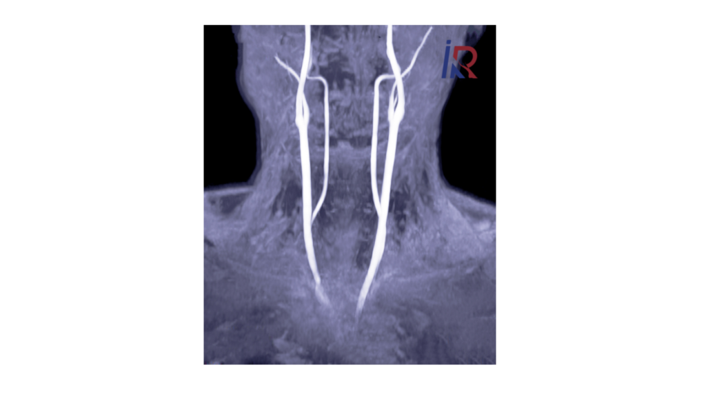
You have two Internal Carotid Arteries (ICA) — one running up each side of your neck. You can actually feel their pulse if you place two fingers gently on either side of your windpipe. These arteries are the main delivery routes carrying oxygen-rich blood from your heart up into your brain. Once inside the skull, each ICA branches out into smaller vessels that supply large portions of the brain responsible for thinking, movement, vision, and sensation.
Think of the two ICAs as the two main highways feeding a busy city. As long as the highway is intact and clear, traffic (blood) flows smoothly. Problems arise when a weak patch develops somewhere along that highway.
So, What Exactly Is an Aneurysm?
An aneurysm is a weak, thin spot in the wall of an artery that balloons outward under the constant pressure of blood flow — much like a thin, worn patch on a bicycle tire bulging outward under air pressure. It fills with blood and sits there, attached to the side or end of the artery, like a small pouch.
As long as it stays small and its wall stays strong, an aneurysm often causes no problems at all and may never be noticed. But two things can go wrong:
- It can grow and start pressing on nearby nerves or brain structures, causing symptoms.
- Its wall can become too thin and rupture (burst), causing sudden bleeding around the brain — a medical emergency.
An Internal Carotid Artery Aneurysm, specifically, is an aneurysm that has formed somewhere along the path of the ICA — and where exactly it forms turns out to matter quite a lot, which brings us to the next section.
Also Read: Internal Carotid Artery Aneurysm Coiling: Treatment, Procedure & Recovery
Where Do These Aneurysms Form? Understanding the ICA’s “Segments” in Simple Terms
Here’s something most simple guides skip, but which genuinely helps you understand your own report: as the Internal Carotid Artery travels from your neck into your brain, doctors divide its journey into several named sections, or segments. Radiologists commonly describe six intracranial segments, sometimes labelled C2 through C7:
- Petrous segment — where the artery first enters the base of the skull, passing through a bony canal
- Lacerum segment — a short section near a skull opening called the foramen lacerum
- Cavernous segment — where the artery runs through a chamber called the cavernous sinus, right beside several important eye-movement nerves
- Clinoid segment — a short transitional section near small bony points at the skull base
- Ophthalmic (paraophthalmic) segment — near where the artery gives off the main artery supplying the eye
- Communicating segment — the final section, near where the ICA joins the brain’s main circulation loop (the Circle of Willis)
Why does this matter to you? Because the symptoms an aneurysm causes often depend heavily on which segment it sits in:
- Aneurysms in the cavernous segment sit close to the nerves controlling eye movement and facial sensation. Studies show over half of patients with cavernous ICA aneurysms present with symptoms from pressure on these nerves — drooping eyelid, double vision, or facial pain — rather than a rupture, because this segment is technically outside the space where bleeding causes the classic “brain bleed” emergency.
- Aneurysms in the ophthalmic segment sit near the artery feeding the eye and can affect vision.
- Aneurysms in the communicating segment sit within the space around the brain itself, meaning rupture here is more likely to cause the classic sudden, severe headache and subarachnoid hemorrhage.
Your doctor will always tell you exactly where your aneurysm sits and what that specific location means for you — but understanding that “location changes everything” helps make sense of why two people with an “ICA aneurysm” can have completely different experiences.
What Causes an Internal Carotid Artery Aneurysm?
There is rarely a single cause. Instead, an aneurysm usually develops gradually, over years, from a combination of factors that weaken the artery wall:
- High blood pressure — long-term pressure stretches and weakens artery walls
- Smoking — one of the strongest known risk factors for both forming and enlarging aneurysms
- Family history — risk is higher if a close relative has had a brain aneurysm
- Atherosclerosis — fatty deposits building up inside the artery wall
- Head injury — trauma can sometimes damage the artery wall directly
- Infection — rarely, an infection can weaken the vessel wall
- Genetic conditions — certain inherited disorders, such as polycystic kidney disease, raise the risk
- Heavy alcohol use and recreational drug use — both raise blood pressure and damage vessels over time
Aneurysms typically become apparent in adults between 40 and 60 years old, and women are diagnosed somewhat more often than men. Because they usually grow silently over many years without any warning, they’re sometimes called a “silent” vascular problem.
Also Read: Vascular Surgeon in Indore | Dr. Alok Kumar Udiya, CARE CHL
Common Symptoms — And Why So Many Aneurysms Are Silent
Unruptured and Symptom-Free
Most small Internal Carotid Artery aneurysms cause no symptoms whatsoever and are discovered by chance — during a scan done for a headache, dizziness, a car accident, or an unrelated check-up. This is actually the most common way an ICA aneurysm is found today.
Unruptured but Symptomatic (Pressing on Nearby Structures)
When an aneurysm grows large enough to press on nearby nerves — especially in the cavernous segment described above — it can cause:
- Pain above or behind one eye
- Drooping of one eyelid
- Double or blurred vision
- A dilated (enlarged) pupil on one side
- Numbness or altered sensation on one side of the face
Ruptured — A Medical Emergency
If the aneurysm’s wall gives way, symptoms appear suddenly and severely:
- A sudden, extremely severe headache — often described as “the worst headache of my life”
- Neck stiffness
- Nausea and vomiting
- Sudden weakness or loss of balance
- Confusion or loss of consciousness
A sudden, unusually severe headache unlike any before it is always a medical emergency and needs immediate hospital care — not a wait-and-see approach.
How Serious Is My Aneurysm? Understanding Rupture Risk
Not every aneurysm carries the same risk, and this is exactly why your doctor looks closely at several factors before recommending a plan:
- Size — larger aneurysms generally carry a higher rupture risk than smaller ones, though small aneurysms can still rupture
- Location — as discussed above, aneurysms within the subarachnoid space carry a different risk profile than those in the cavernous segment
- Shape — irregular shapes or small outpouchings (“daughter sacs”) on the aneurysm wall can indicate higher risk
- Growth over time — an aneurysm that is growing on repeat scans is treated more urgently than one that has stayed stable for years
- Your personal risk factors — blood pressure control, smoking status, and family history all factor into the overall picture
This is why two people with a similarly sized aneurysm may receive different recommendations — one for careful monitoring, another for active treatment. It is a genuinely individualised decision, not a one-size-fits-all rule.
How Is an Internal Carotid Artery Aneurysm Diagnosed?
Once an aneurysm is suspected, your doctor will typically use one or more of the following:
- CT Scan of the brain — a fast scan, especially important if a rupture is suspected, as it can show bleeding
- CT Angiography (CTA) — uses an injected dye to map the blood vessels in detail
- MRI and MR Angiography (MRA) — provides clear pictures of the brain and its vessels without radiation
- Digital Subtraction Angiography (DSA) — a live, moving X-ray of the blood vessels using a thin catheter and dye; considered the most detailed and accurate test, and also used to guide treatment when needed
At CARE CHL Hospital, Indore, all of these imaging tools are available under one roof, allowing a fast, accurate diagnosis and a clear next step.
Also Read: Gangrene Treatment in Indore: Advanced Care & Trusted Specialists
Treatment Options — In Simple Terms
Broadly, there are two paths for treating an Internal Carotid Artery aneurysm that needs active management:
Surgical clipping — traditional open brain surgery where a surgeon places a small metal clip at the base of the aneurysm to seal it off. This remains a good option for certain locations, such as some aneurysms near the eye artery (ophthalmic segment), but involves opening the skull, a longer hospital stay, and a longer recovery.
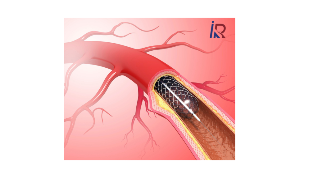
Endovascular coiling — the modern, minimally invasive approach, where soft platinum coils are guided into the aneurysm through a thin catheter inserted in the wrist or groin, sealing it off from the inside without opening the skull. Increasingly, techniques like balloon-assisted coiling, stent-assisted coiling, and flow diversion are also used for more complex aneurysm shapes.
Watchful monitoring — for some small, stable, low-risk aneurysms found incidentally, your doctor may recommend regular follow-up imaging rather than immediate treatment, especially if the risks of treatment outweigh the current risk of rupture.
This guide intentionally keeps the treatment section brief — if you’d like a full, step-by-step walkthrough of exactly how the coiling procedure works, what to expect on the day, and recovery timelines, our companion guide covers Internal Carotid Artery Aneurysm Coiling in complete detail.
Meet Dr. Alok Kumar Udiya — Interventional Radiologist, Indore
If you’ve been told you or a loved one has an Internal Carotid Artery aneurysm, the specialist you choose to guide you matters enormously.
Dr. Alok Kumar Udiya is a Senior Consultant Interventional Radiologist at CARE CHL Hospital, Indore, with training that specifically covers brain aneurysm care:
- MBBS — M.G.M. Medical College, Indore
- MD (Radiodiagnosis) — Lady Hardinge Medical College, Delhi University
- PDCC (Neuro-Intervention) — Sanjay Gandhi Post Graduate Institute of Medical Sciences (SGPGIMS), Lucknow
- PDCC (Hepatobiliary Interventional Radiology) — Institute of Liver and Biliary Sciences (ILBS), New Delhi
- FINR — Fellowship in Interventional Neuroradiology, University of Zurich, Switzerland, trained directly under Professor Anton Valavanis, specifically in the endovascular management of stroke and brain aneurysms
- EBIR — European Board of Interventional Radiology certified
He is a life member of the Indian Radiological and Imaging Association (IRIA) and the Indian Society of Vascular and Interventional Radiology (ISVIR), with peer-reviewed publications on aneurysm coiling techniques and related neurovascular topics. Patients and families consistently describe his approach as calm and clear — explaining a frightening diagnosis in words that make sense, before ever discussing treatment.
Learn more on the About Us page or explore the full range of Neuro Interventions offered.
Why Choose Intervention Radiology Indore at CARE CHL Hospital (LIG Square)?
Patients with a brain aneurysm no longer need to travel to Delhi, Mumbai, or Ahmedabad for advanced care. Intervention Radiology Indore operates from CARE CHL Hospital, a NABH-accredited, 225-bed multi-specialty hospital serving central India since 2001, offering:
- A dedicated neuro-interventional suite with Digital Subtraction Angiography (DSA) for precise aneurysm mapping
- Advanced CT and MR imaging on-site for fast, accurate diagnosis
- A specialist trained in Zurich specifically in aneurysm and stroke endovascular care
- A multidisciplinary team of neurologists, neurosurgeons, and critical care specialists working together on complex cases
- Consultations available in Hindi and English
Clinic Locations, Timings & How to Book
CARE CHL Hospital (near LIG Square) 📍 AB Road, near L.I.G. Square, RSS Nagar, Indore, Madhya Pradesh – 452008 🕙 OPD consultation hours: 10:00 AM – 5:00 PM
Evening Clinic — Panama Tower 📍 403, Panama Tower, Geeta Bhawan Square, near Crown Palace, South Tukoganj, Indore, Madhya Pradesh – 452001 🕕 Consultation hours: 6:00 PM – 8:00 PM
📞 Call/WhatsApp: +91 99993 78980 ✉️ Email: dralokudiya@gmail.com 🌐 Website: interventionradiologyindore.com
The LIG Square location on AB Road is a well-known landmark, making the hospital easy to reach from anywhere in Indore, Dewas, Ujjain, or the wider Malwa region.
Myths and Facts About Internal Carotid Artery Aneurysms
Myth: Every aneurysm needs immediate surgery. Fact: Many small, stable, incidentally found aneurysms are simply monitored with periodic scans rather than treated right away.
Myth: If it hasn’t burst yet, it never will. Fact: Risk depends on size, shape, location, and growth over time — not just how long it has already existed without rupturing.
Myth: An aneurysm always causes warning symptoms before it ruptures. Fact: Most unruptured aneurysms are completely silent. Absence of symptoms does not mean absence of risk.
Myth: All aneurysm treatment means open brain surgery. Fact: The majority of Internal Carotid Artery aneurysms today can be treated with minimally invasive endovascular coiling, without opening the skull.
Myth: Aneurysms are extremely rare. Fact: Internal Carotid Artery aneurysms are among the more commonly diagnosed types of brain aneurysms, especially now that brain imaging is more widely used.
Frequently Asked Questions
What is an Internal Carotid Artery Aneurysm in simple words?
It’s a weak, bulging spot — like a small balloon — on the wall of one of the two main arteries carrying blood to your brain. It can stay silent for years or, less commonly, grow, press on nearby nerves, or rupture.
Is an Internal Carotid Artery Aneurysm always dangerous?
Not always. Many small, stable aneurysms found incidentally on a scan carry a low risk and are simply monitored. Risk depends on the aneurysm’s size, shape, location, and whether it is growing.
What are the first warning signs of a problem?
Symptoms vary by location — they can include pain behind the eye, a drooping eyelid, or double vision if the aneurysm is pressing on a nerve. A sudden, extremely severe headache is the hallmark warning sign of a rupture and is always a medical emergency.
How is it diagnosed?
Through imaging such as a CT scan, CT angiography, MRI/MRA, or a Digital Subtraction Angiogram (DSA) — the most detailed test, also used to guide treatment.
Does every Internal Carotid Artery Aneurysm need coiling or surgery?
No. Small, stable, low-risk aneurysms are often simply monitored with periodic imaging. When treatment is needed, endovascular coiling is the preferred approach for most locations today.
Who should I consult for an Internal Carotid Artery Aneurysm in Indore?
Dr. Alok Kumar Udiya, Interventional Radiologist at CARE CHL Hospital, near LIG Square, Indore, specialises in the diagnosis and endovascular treatment of brain aneurysms. Call or WhatsApp +91 99993 78980 to book a consultation.
Conclusion
An Internal Carotid Artery Aneurysm can feel like an alarming, confusing diagnosis when you first hear the word. But as this guide has hopefully shown, it is also a condition that modern medicine understands extremely well — where it forms, why it forms, what it might cause, and how to treat it safely when treatment is truly needed.
If you or a loved one has recently been told about an aneurysm, the most useful next step isn’t panic — it’s a clear, expert conversation with a specialist who can look at your specific scan, your specific location, and your specific risk factors, and explain exactly what it means for you.
Dr. Alok Kumar Udiya at CARE CHL Hospital, LIG Square, brings internationally trained neurovascular expertise to patients across Indore and central India — ready to answer your questions and guide you toward the safest path forward.
Book Your Consultation Today
🚨 Sudden, severe headache or other emergency symptoms? Go to the nearest emergency room immediately.
📍 CARE CHL Hospital, AB Road, near LIG Square, RSS Nagar, Indore, MP 452008 (OPD: 10 AM-5 PM) 📍 Evening Clinic, 403 Panama Tower, Geeta Bhawan Square, Indore, MP 452001 (6 PM-8 PM) 📞 +91 99993 78980 🌐 interventionradiologyindore.com 👨⚕️ Dr. Alok Kumar Udiya — Interventional Radiologist, Indore
Have a scan report mentioning an Internal Carotid Artery Aneurysm? Book a consultation with Intervention Radiology Indore for a clear, expert opinion.
Disclaimer: This blog is intended for educational purposes only and does not constitute medical advice, diagnosis, or emergency guidance. A sudden, severe headache or other emergency symptom requires immediate emergency care. Please consult a qualified medical professional for diagnosis and treatment of any health condition.